Article Text

Statistics from Altmetric.com
“It seems probable that this study covers the period of practical extinction of empyema as an important disease.” Lionakis B et al, J Pediatr 1958.
1. SEARCH METHODOLOGY
1.1 Structure of the guideline
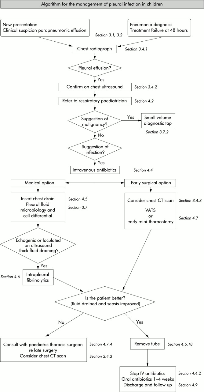
The format follows that used for the BTS guidelines on the management of pleural disease in adults.1 At the start there is a summary table of the abstracted bullet points from each section. Following that is an algorithm summarising the management of pleural infection in children (fig 1). Each section starts with bulleted points of key recommendations using the revised SIGN grading system (table 1) available on http://www.sign.ac.uk/guidelines/fulltext/50/section6.html. Beneath each set of bullet points is a short paragraph detailing the referenced literature and the rationale behind the recommendations. The primary source literature has been individually graded for its methodology and the grading is given alongside each reference using the revised SIGN levels of evidence (table 2).
Revised SIGN grading system: grades of recommendation
Revised SIGN grading system: levels of evidence
Algorithm for the management of pleural infection in children.
1.2 Methodology for generation of the guidelines
The initial literature search was carried out by the Library of the National Heart Lung Institute, Imperial College London. Further searches were then carried out by members of the working group who concentrated on their own topics. Details of the search strategy are given in Appendix 1.
Each section of the guideline was researched and drafted by a subgroup of the Paediatric Pleural Diseases Subcommittee (itself a subcommittee of the BTS Standards of Care Committee). Publications were rated according to the SIGN criteria for the calibre of the methodology of the research to give levels of evidence (table 2). Tables of evidence were then produced before writing the guideline sections using the SIGN grades of recommendations (table 1). Once all parts were merged into one document, the whole group then met to discuss the first draft before redrafting took place. This draft was based, where possible, on the published evidence but this was then combined with clinical expertise as required. The resulting draft is therefore a blend of published evidence and clinical experience. This was sent to a group of specialist reviewers listed in the Acknowledgements.
The manuscript was then amended in the light of their comments and the document was reviewed by the BTS Standards of Care Committee following which a further drafting took place. The Quality of Practice Committee of the Royal College of Paediatrics and Child Health also reviewed this draft. After final approval from this Committee, the guidelines were submitted for blind peer review and publication.
1.3 Conflict of interest
All the members of the Guideline Committee submitted a written record of possible conflicts of interest to the Standards of Care Committee of the BTS. There were none. These are available for inspection on request from the Chairman of this Committee.
1.4 Acknowledgements
Funding for literature searches, photocopying and travel to the guideline meeting was kindly provided by the British Thoracic Society. The authors thank the library staff at the National Heart Lung Institute and John Vandridge-Ames at the Royal College of Radiologists for additional searches, Dr Juliet Hale (Consultant Paediatric Oncologist, Royal Victoria Hospital, Newcastle upon Tyne) for advice on malignant effusions, Dr Elizabeth Haxby (Consultant Anaesthetist and Lead Clinician for Clinical Risk, Royal Brompton Hospital, London) for advice on anaesthesia and sedation, Dr Jon Smith (Consultant in Paediatric Cardiothoracic Anaesthesia and Intensive Care, Freeman Hospital, Newcastle upon Tyne) for advice on analgesia, Dr Ravi Jayaram (Specialist Registrar, Royal Brompton Hospital, London) for advice on Appendix 3, and Jayne Wellington for the mountain of photocopying.
The following acted as specialist reviewers: Dr Robert Dinwiddie (Consultant in Paediatric Respiratory Medicine, Great Ormond Street Hospital for Children, London), Dr Iolo Doull (Consultant in Paediatric Respiratory Medicine, University Hospital of Wales, Cardiff), Mr Peter Goldstraw (Consultant Thoracic Surgeon, Royal Brompton Hospital, London), Dr Robert Primhak (Senior Lecturer and Honorary Consultant in Paediatric Respiratory Medicine, Sheffield Children’s Hospital), Dr Paul Seddon (Consultant Paediatrician with an interest in Respiratory Medicine, Royal Alexandra Hospital for Sick Children, Brighton).
The authors also thank the Quality of Practice Committee of the Royal College of Paediatrics and Child Health for reviewing the guidelines and particularly for their comments on methodology.
2. INTRODUCTION
2.1 The need for paediatric guidelines
Although still relatively uncommon, it seems that pleural infections have become more prevalent in the UK2,3 and USA.4,5 More cases are being seen in paediatric respiratory centres, and with fewer chest drains being inserted in district general hospitals, they are often seen at an earlier stage by respiratory paediatricians. Empyemas are a significant cause of morbidity but, fortunately, not mortality in children, and at times can be a therapeutic challenge. Despite this, in the UK there is little consensus over management among respiratory paediatricians and thoracic surgeons. Part of the problem has been the lack of evidence from paediatric trials, and it is inappropriate simply to extrapolate adult data to children. There are differences between adult and paediatric pleural infections. The principal one is that, since it is rare for children to have an underlying lung disease, the final outcome is almost always excellent. This is in contrast to the disease in adults where empyema is a cause of significant morbidity with 40% of patients requiring pleural surgery due to failed catheter drainage.6 Furthermore, adult empyema carries a 20% mortality rate7 which is related to co-morbidity (for example, malignancy, immunodeficiency, prolonged hospital stay and nosocomially acquired infection). With the publication of the BTS guidelines for the management of pleural disease (in adults),1 it seemed appropriate to produce some for children.
This guideline has assessed available evidence and attempted to gauge consensus opinion where evidence is unavailable. The lack of paediatric data, in particular from randomised controlled trials, is reflected in the grading of levels of evidence and recommendations in this document. Although there are many grade D recommendations, some of these are safe current practice based on common sense but, since they have never been subjected to a randomised controlled trial, they remain a grade D. An example would be the recommendation to send pleural fluid for bacterial culture. Clearly a D label should not necessarily undermine the significance of the recommendation. For some issues, evidence from adult practice has been assessed and referred to if it seemed applicable to children. It is hoped that these guidelines will facilitate dissemination of evidence, standardisation of patient care, and reduce the morbidity in these patients.
2.2 Epidemiology
Parapneumonic effusion and empyema have an incidence of 3.3 per 100 000 children.4 It has been suggested that the incidence of childhood empyema increased in the UK in the mid to late 1990s,2,3 although this is not a universal finding.8 It is not clear whether this is related to different referral patterns, changes of antibiotic usage in primary care, or whether it was a genuine increase in disease incidence. Parapneumonic effusions and empyema are more common in boys than girls and are more frequently encountered in infants and young children.9 They are also more common in winter and spring,9 presumably due to their infective origin.
2.3 Definition and staging
The definitions of parapneumonic effusion (pleural fluid collection in association with underlying pneumonia) and empyema (the presence of pus in the pleural space) are best considered by reviewing the staging of pleural fluid associated with infection. Pleural infection is a continuum but classically it has been divided into three stages:10
-
Exudative: the inflammatory process associated with the underlying pneumonia leads to the accumulation of clear fluid with a low white cell count within the pleural cavity (simple parapneumonic effusion).
-
Fibropurulent: there is deposition of fibrin in the pleural space leading to septation and the formation of loculations. There is an increase in white cells, with the fluid thickening (complicated parapneumonic effusion) and eventually becoming overt pus (empyema). The presence of septations (fibrinous strands within the pleural fluid) does not necessarily mean the fluid does not flow freely, although separate loculations will not communicate with each other.11
-
Organisational: fibroblasts infiltrate the pleural cavity, and the thin intrapleural membranes are reorganised to become thick and non-elastic (the “peel”). These solid fibrous pleural peels may prevent lung re-expansion (“trapped lung”), impair lung function, and create a persistent pleural space with ongoing potential for infection. At this stage spontaneous healing may occur or a chronic empyema may develop.
Further complications are uncommon in children but may include bronchopleural fistula, lung abscess, or even perforation through the chest wall (empyema necessitatis).
Abstracted bullet points
Clinical picture
-
All children with parapneumonic effusion or empyema should be admitted to hospital. [D]
-
If a child remains pyrexial or unwell 48 hours after admission for pneumonia, parapneumonic effusion/empyema must be excluded. [D]
Diagnostic imaging
-
Posteroanterior or anteroposterior radiographs should be taken; there is no role for a routine lateral radiograph. [D]
-
Ultrasound must be used to confirm the presence of a pleural fluid collection. [D]
-
Ultrasound should be used to guide thoracocentesis or drain placement. [C]
-
Chest CT scans should not be performed routinely. [D]
Diagnostic microbiology
-
Blood cultures should be performed in all patients with parapneumonic effusion. [D]
-
When available, sputum should be sent for bacterial culture. [D]
Diagnostic analysis of pleural fluid
-
Pleural fluid must be sent for microbiological analysis including Gram stain and bacterial culture. [C]
-
Aspirated pleural fluid should be sent for differential cell count. [D]
-
Tuberculosis and malignancy must be excluded in the presence of pleural lymphocytosis. [C]
-
If there is any indication the effusion is not secondary to infection, consider an initial small volume diagnostic tap for cytological analysis, avoiding general anaesthesia/sedation whenever possible. [D]
-
Biochemical analysis of pleural fluid is unnecessary in the management of uncomplicated parapneumonic effusions/empyema. [D]
Diagnostic bronchoscopy
-
There is no indication for flexible bronchoscopy and it is not routinely recommended. [D]
Referral to tertiary centre
-
A respiratory paediatrician should be involved early in the care of all patients requiring chest tube drainage for a pleural infection. [D]
Conservative management (antibiotics ± simple drainage)
-
Effusions which are enlarging and/or compromising respiratory function should not be managed by antibiotics alone. [D]
-
Give consideration to early active treatment as conservative treatment results in prolonged duration of illness and hospital stay. [D]
Repeated thoracocentesis
-
If a child has significant pleural infection, a drain should be inserted at the outset and repeated taps are not recommended. [D]
Antibiotics
-
All cases should be treated with intravenous antibiotics and must include cover for Streptococcus pneumoniae. [D]
-
Broader spectrum cover is required for hospital acquired infections, as well as those secondary to surgery, trauma, and aspiration. [D]
-
Where possible, antibiotic choice should be guided by microbiology results. [B]
-
Oral antibiotics should be given at discharge for 1–4 weeks, but longer if there is residual disease. [D]
Chest drains
-
Chest drains should be inserted by adequately trained personnel to reduce the risk of complications. [C]
-
A suitable assistant and trained nurse must be available. [D]
-
Routine measurement of the platelet count and clotting studies are only recommended in patients with known risk factors. [D]
-
Where possible, any coagulopathy or platelet defect should be corrected before chest drain insertion. [D]
-
Ultrasound should be used to guide thoracocentesis or drain placement. [C]
-
If general anaesthesia is not being used, intravenous sedation should only be given by those trained in the use of conscious sedation, airway management and resuscitation of children, using full monitoring equipment. [D]
-
Small bore percutaneous drains should be inserted at the optimum site suggested by chest ultrasound. [C]
-
Large bore surgical drains should also be inserted at the optimum site suggested by ultrasound, but preferentially placed in the mid axillary line through the “safe triangle”. [D]
-
Since there is no evidence that large bore chest drains confer any advantage, small drains (including pigtail catheters) should be used whenever possible to minimise patient discomfort. [C]
-
Neither substantial force nor a trocar should ever be used to insert a drain. [D]
-
A chest radiograph should be performed after insertion of a chest drain. [D]
-
All chest tubes should be connected to a unidirectional flow drainage system (such as an underwater seal bottle) which must be kept below the level of the patient’s chest at all times. [D]
-
Appropriately trained nursing staff must supervise the use of chest drain suction. [D]
-
A bubbling chest drain should never be clamped. [D]
-
A clamped drain should be immediately unclamped and medical advice sought if a patient complains of breathlessness or chest pain. [D]
-
The drain should be clamped for 1 hour once 10 ml/kg are initially removed. [D]
-
Patients with chest drains should be managed on specialist wards by staff trained in chest drain management. [D]
-
When there is a sudden cessation of fluid draining, the drain must be checked for obstruction (blockage or kinking) by flushing. [D]
-
The drain should be removed once there is clinical resolution. [D]
-
A drain that cannot be unblocked should be removed and replaced if significant pleural fluid remains. [D]
Intrapleural fibrinolytics
-
Intrapleural fibrinolytics shorten hospital stay and are recommended for any complicated parapneumonic effusion (thick fluid with loculations) or empyema (overt pus). [B]
-
There is no evidence that any of the three fibrinolytics are more effective than the others, but only urokinase has been studied in a randomised controlled trial in children so is recommended. [B]
-
Urokinase should be given twice daily for 3 days (6 doses in total) using 40 000 units in 40 ml 0.9% saline for children weighing 10 kg or above, and 10 000 units in 10 ml 0.9% saline for children weighing under 10 kg. [B]
Surgery
-
Failure of chest tube drainage, antibiotics, and fibrinolytics should prompt early discussion with a thoracic surgeon. [D]
-
Patients should be considered for surgical treatment if they have persisting sepsis in association with a persistent pleural collection, despite chest tube drainage and antibiotics. [D]
-
Organised empyema in a symptomatic child may require formal thoracotomy and decortication. [D]
-
A lung abscess coexisting with an empyema should not normally be surgically drained. [D]
Other management
-
Antipyretics should be given. [D]
-
Analgesia is important to keep the child comfortable, particularly in the presence of a chest drain. [D]
-
Chest physiotherapy is not beneficial and should not be performed in children with empyema. [D]
-
Early mobilisation and exercise is recommended. [D]
-
Secondary thrombocytosis (platelet count >500 × 109/l) is common but benign; antiplatelet therapy is not necessary. [D]
-
Secondary scoliosis noted on the chest radiograph is common but transient; no specific treatment is required but resolution must be confirmed. [D]
Follow up
-
Children should be followed up after discharge until they have recovered completely and their chest radiograph has returned to near normal. [D]
-
Underlying diagnoses—for example, immunodeficiency, cystic fibrosis—may need to be considered. [D]
2.4 Pathophysiology
The pleural space normally contains 0.3 ml/kg body weight of pleural fluid.12 There is a continuous circulation of this fluid and the lymphatic vessels can cope with several hundred millilitres of extra fluid per 24 hours.13 However, an imbalance between pleural fluid formation and drainage will result in a pleural effusion. In health, pleural fluid contains a small number of cells (mainly mesothelial cells, macrophages, lymphocytes) with a low protein concentration (0.1 g/l), as well as large molecular weight proteins such as lactate dehydrogenase (LDH). Compared with the serum, the pleural fluid has higher levels of bicarbonate, lower levels of sodium, and similar levels of glucose.6
These parameters are altered when disease processes such as infection affect the adjacent lung or vascular tissue and activate an immune response and pleural inflammation. Increased vascular permeability allows migration of inflammatory cells (neutrophils, lymphocytes, and eosinophils) into the pleural space. The process is mediated by a number of cytokines—such as interleukin (IL)-1, IL-6, IL-8, tumour necrosis factor (TNF)-α, and platelet activating factor—released by mesothelial cells lining the pleural space.12 The result is the exudative stage of a pleural effusion. This progresses to the fibropurulent stage due to increased fluid accumulation and bacterial invasion across the damaged epithelium.6 Neutrophil migration occurs as well as activation of the coagulation cascade leading to procoagulant activity and decreased fibrinolysis.14 Deposition of fibrin in the pleural space then leads to septation or loculation. The pleural fluid pH and glucose level falls while LDH levels increase.15
2.5 Aetiology
In a previously well child, pleural effusions are usually secondary to acute bacterial pneumonia9 and less often due to chronic infections such as pulmonary tuberculosis.16 When associated with infection, effusions are usually unilateral and bilateral empyemas are unusual, except in one large Turkish series of 515 children where 5% were bilateral.17 Bilateral effusions may indicate tuberculosis or a parasitic infection.18 The rate of parapneumonic effusion complicating pneumonia is said to be 1%,19 although it has been suggested that effusions may be found in up to 40% of adult cases admitted to hospital.10 The prevalence of small parapneumonic effusions is difficult to estimate (and often undetected), and they are unlikely to be reported in case series. Other infections such as lung abscess and chronic suppurative conditions such as bronchiectasis may also produce pleural effusion.9 Predisposing causes include immunodeficiencies, aspiration, post-surgery and trauma.
Pleural effusions are not always secondary to infection and may be genuinely sterile. Rarely, an effusion is the presenting sign of an underlying malignancy in a child who was well before the symptoms related to the effusion. Many of the other secondary causes of pleural effusion will be in children with a known underlying condition such as congenital heart disease, renal disease, connective tissue disorders, and trauma which includes post-cardiothoracic surgery. There are several published case series reporting causes of effusions in children but the proportion of non-infective causes is largely dependent on the referral base and case mix in the particular hospital.9,19–21
2.6 Microbiology
The epidemiology has altered significantly over the last 70 years with the discovery of new antibiotics that have different spectra of activity for use in pneumonia. The reported rate of identifying an infectious organism from pleural fluid varies markedly, from 8% to 76%.9,19,21 Precise information is unavailable since much of the historical data is unhelpful due to differences in definitions and inclusion/exclusion criteria. This is further hampered by different pleural fluid sampling rates as well as different culture and identification techniques. Furthermore, in present day practice, pleural fluid culture is often sterile because of antibiotics used before obtaining a pleural fluid sample. In the recent multicentre UK study only 17% of cases were culture positive.22 Even using newer molecular techniques—for example, pneumococcal or broad range 16S polymerase chain reaction (PCR)—an aetiological agent was only detected in about 75% of culture negative cases, although this does represent an improvement.23,24
2.6.1 Acute bacterial infection
In the pre-antibiotic era, Streptococcus pneumoniae was the major pathogen recovered from pleural fluid, followed by β-haemolytic streptococci (probably Streptococcus pyogenes) and Staphylococcus aureus.25,26 With the introduction of sulphonamides and then penicillin, the incidence of S pneumoniae and S pyogenes was markedly reduced and the relative proportion of S aureus increased, especially in the late 1950s as the rate of penicillin resistant S aureus began to increase.25S aureus was particularly evident in the first 6 months of life, and overall accounted for 29%9 to 63%27 of cases. There have also been reports of empyema due to methicillin-resistant S aureus in children.28,29
Following the introduction of penicillinase stable penicillins and other antistaphylococcal agents, the relative proportion of empyema due to S pneumoniae has increased once more. Currently it seems to be emerging as the predominant pathogen in childhood empyema, although this is not always reflected in culture results as many are culture negative.3,30–32 Nevertheless, S pneumoniae was the principal organism in three recent case series from the USA,4,29,33 and the majority of culture negative cases in two UK series have been shown to be S pneumoniae by molecular techniques.23,24 In the Newcastle study, evidence of S pneumoniae was found in 75% culture negative pleural fluid samples by PCR methods as well as latex agglutination testing for pneumococcal antigen;24 53% of these were capsular serotype 1 and all were penicillin sensitive.
Other bacteria include S pyogenes,19,34Haemophilus influenzae type b,21Mycoplasma pneumoniae,35,36Pseudomonas aeruginosa,27,37 and other streptococcal species (including viridans streptococci38 and streptococci of Lancefield group F39). Rarer bacterial organisms isolated include Klebsiella,40Enterobacter,37Proteus species,37Salmonella,41 and Yersinia.42 Anaerobic organisms such as Bacteroides species and Peptostreptococcus are rarely isolated in children but may be associated with aspiration pneumonia or foreign bodies,12,43 as may Streptococcus milleri;22 they must always be considered in children with delayed neurodevelopment. Disseminated Fusobacteriumnecrophorum infection (Lemierre syndrome) is a potentially fatal condition which typically follows a severe pharyngitis and may be seen in older children (and young adults); although rare, it seems to be increasing in incidence.44
The bacterial aetiological profile differs in developing countries with S aureus being the predominant pathogen, especially during the hot and humid months when staphylococcal skin infections are more prevalent.17,45 There has been a decline in culture positive S pneumoniae, probably because of prior antibiotic use.45 Various Gram negative organisms—for example, Enterobacteriaceae such as Klebsiella spp and Pseudomonas aeruginosa—are also more common than in the UK; they are not limited to infants and may be associated with protein energy malnutrition.27,37,45,46
2.6.2 Mycoplasma, Legionella and viruses
Pleural effusion is reported in association with mycoplasma infection although empyema is rare.47 Mycoplasma serology, when performed, suggests involvement in some cases30,36 but most series do not report serology results and paired samples may not have been taken. Legionella pneumophila48 and primary viral pneumonia49 may also be associated with pleural effusion but the contribution of these agents to pleural empyema is not accurately known as few studies report adequate investigations of all cases. Besides, a viral infection may simply precede a secondary bacterial infection which then causes the empyema. Certainly adenovirus36,49 and influenza virus35 can cause effusions, but they are rarely large.
2.6.3 Mycobacterial infection
Tuberculous empyema can result from progressive pulmonary tuberculosis. It has been reported to account for up to 6% of all empyema cases worldwide,6 but with aggressive modern antituberculous chemotherapy it is seldom seen in the UK.12
2.6.4 Other organisms
Fungal causes are usually nosocomial in origin50,51 or, in the case of the rare Histoplasma infection, follow exposure.52,53 Finally, there is a single case report of Entamoeba histolytica.54
2.7 Clinical picture
-
All children with parapneumonic effusion or empyema should be admitted to hospital. [D]
-
If a child remains pyrexial or unwell 48 hours after admission for pneumonia, parapneumonic effusion/empyema must be excluded. [D]
There are two common patterns of presentation. In the first, the child has classic symptoms of pneumonia—for example, fever, cough, breathlessness, exercise intolerance, poor appetite, abdominal pain, fetor oris (halitosis), lethargy and malaise.55 However, in the presence of an effusion they are often more unwell than with simple pneumonia alone. They may have pleuritic chest pain and may lie on the affected side to splint the involved hemithorax and provide temporary analgesia.12 On examination a pleural effusion is suggested by unilateral signs of decreased chest expansion, dullness to percussion, reduced or absent breath sounds, and scoliosis. There may also be cyanosis due to ventilation-perfusion mismatch. The effusion is often obvious on the initial chest radiograph. All children with parapneumonic effusion or empyema should be admitted to hospital.
The second scenario is of the child who has been diagnosed with pneumonia but does not respond to the usual and appropriate treatment. We would reiterate the recommendations from BTS guidelines for the management of community acquired pneumonia in childhood55 that, if a child remains pyrexial or unwell 48 hours after admission with pneumonia, re-evaluation is necessary with consideration given to possible complications. Careful clinical examination and a repeat chest radiograph are warranted.
2.8 Outcome and prognosis
The prognosis in children with empyema is usually very good. Follow up studies have shown that, despite the heterogeneity of treatment approaches, the majority of children make a complete recovery and their lung function returns to normal.56–63 Other studies have shown minor abnormalities in lung function of both a restrictive64,65 and obstructive nature,61 but the children were still asymptomatic with normal exercise tolerance.61,64,65 The chest radiograph returns to normal in the majority of children (60–83%) by 3 months, in over 90% by 6 months, and in all by 18 months.30,62
3. DIAGNOSIS
3.1 Clinical history
The child with a parapneumonic effusion/empyema usually presents with classic symptoms of pneumonia (cough, dyspnoea, fever, malaise, loss of appetite), although perhaps they are more unwell than usual and may have pleuritic chest pain. Infection in the lower lobes may present with abdominal pain. In those already diagnosed with pneumonia, a spiking fever and lack of improvement after 48 hours of antibiotic treatment may signal the presence of an effusion. Antibiotic history is important and underlying rarer conditions (such as tuberculosis, immunodeficiency, inhaled foreign body, and malignancy) must be considered.
3.2 Physical examination
A pleural effusion is suggested by unilateral signs of decreased chest expansion, dullness to percussion, and reduced or absent breath sounds. The assessment of severity is the same as that for any childhood pneumonia (table 3), but measurement of oxygen saturation (Spao2) is particularly important with levels below 92% indicating severe disease.55 Examination should also include assessment of the child’s state of hydration, their height and weight, the presence of a scoliosis, and any underlying disorders.
Clinical severity assessment55
3.3 Initial investigations
Initial investigations for a suspected parapneumonic effusion are listed in box 1.
Box 1 Initial investigations for suspected parapneumonic effusion
-
Chest radiograph
-
Ultrasound scan of chest
-
Blood culture (including anaerobic bottle)
-
Sputum culture (if available)
-
Antistreptolysin O titre (ASOT)
-
Full blood count (for anaemia, white count with differential, platelet count)
-
Electrolytes (to detect inappropriate ADH syndrome)
-
Serum albumin (often low)
-
C-reactive protein (some regard this as a useful marker of progress)
3.4 Imaging
3.4.1 Chest radiograph
-
Posteroanterior or anteroposterior radiographs should be taken; there is no role for a routine lateral radiograph. [D]
Obliteration of the costophrenic angle is the earliest sign of a pleural effusion, and a rim of fluid may be seen ascending the lateral chest wall (meniscus sign) on a posteroanterior or anteroposterior radiograph. If the film is taken when a (younger) child is supine, the appearance can be of a homogeneous increase in opacity over the whole lung field without blunting of the costophrenic angle or a classic pleural based shadow.66 When there is a “white out” it is not always possible to differentiate solid underlying severe lung collapse/consolidation from a large effusion. Radiographs alone cannot differentiate an empyema from a parapneumonic effusion.66 A lateral chest radiograph rarely adds anything extra, although can sometimes be helpful in differentiating pleural from intrapulmonary shadows—for example, air in the intrapleural space v an intrapulmonary abscess cavity. Finally, any scoliosis can be detected on a plain chest radiograph.
3.4.2 Ultrasound scan of chest
-
Ultrasound must be used to confirm the presence of a pleural fluid collection. [D]
-
Ultrasound should be used to guide thoracocentesis or drain placement. [C]
Chest ultrasonography can detect the presence of fluid in the pleural space, so is particularly useful when there is a “white out” on the chest radiograph.6 Although ultrasound cannot reliably establish the stage of pleural infection,11 it can estimate the size of the effusion, differentiate free from loculated pleural fluid, and determine the echogenicity of the fluid.66 Ultrasound may also demonstrate pleural thickening and assist in the diagnosis of effusion secondary to tuberculosis (for example, the presence of diffuse small nodules on the pleural surface).67 Finally, it can be used to guide chest drain insertion or thoracocentesis with the radiologist or radiographer marking the optimum site for drainage on the skin.68–71 Ultrasound can conveniently be carried out at the bedside with modern portable units.
3.4.3 Is a CT scan necessary in addition to ultrasound?
-
Chest CT scans should not be performed routinely. [D]
Radiation from a CT chest scan can be high (depending on several factors including the machine, scanning technique, and size of the child), ranging from up to 400 chest radiograph equivalents to as few as 20. There has been little research on the use of ultrasound and CT scanning in paediatric empyema. However, as discussed in section 3.4.2, ultrasound can confirm the presence of pleural fluid (differentiating it from pulmonary infiltrates) so is critical in the diagnosis of parapneumonic effusion/empyema. Although ultrasound cannot usually identify the stage of the pleural effusion,11 a study of 320 adults and some children showed that it might sometimes help to distinguish exudative pleural effusions from transudates.72 The exudates appeared as complex effusions or homogeneously echogenic effusions on ultrasound and these were due either to empyema or haemorrhage. Fibrinous septations are better visualised using ultrasound than CT scans. Ultrasound has also been shown to be good at distinguishing fluid from solid material in the pleural space.73 It will not predict those patients who will fail with chest drain and fibrinolytics alone and subsequently require surgery.11 Ultrasound scanning is now readily available and is the preferred investigation in children, especially as no sedation is necessary and it involves no radiation. It enables the exact location of any fluid collection to be determined and allows guided diagnostic aspiration if required.70,71 Ultrasound is sufficient in the majority of paediatric cases.
In a study of 30 children CT scanning was not helpful in differentiating empyema from parapneumonic effusion.74 Furthermore, in a review of ultrasound and CT scanning in a group of 50 adults with parapneumonic effusion requiring drainage, neither technique reliably identified the stage of the pleural effusion, although pleural thickness on the CT scan was greater in those with frankly purulent effusions.11 CT scanning of the chest with contrast enhancement assists in delineating loculated pleural fluid and can also detect airway or parenchymal lung abnormalities such as endobronchial obstruction or a lung abscess, as well as helping with mediastinal pathology.75,76 While unnecessary for most cases of paediatric empyema, it has a role in complicated cases (including initial failure to aspirate pleural fluid and failing medical management) and particularly in immunocompromised children where a CT scan could reveal other serious clinical problems. Many surgeons will require a CT scan before surgery (either open thoracotomy or thoracoscopy) to delineate the anatomy further and to check for an intrapulmonary abscess.
3.5 Blood tests
Are blood tests helpful in the investigation or management of parapneumonic effusions/empyema?
-
Blood cultures should be performed in all patients with parapneumonic effusion. [D]
3.5.1 Blood cultures
In the BTS guidelines for community acquired pneumonia (CAP) in children it is recommended that blood cultures should be performed in all children suspected of having bacterial pneumonia.55 A recent large retrospective case series of 540 children in the USA with CAP, 153 of whom went on to develop an empyema, confirms that this is worthwhile.5 Blood cultures were positive in 15/153 (10%) with empyema and 25/387 (6.4%) of those with pneumonia alone. Another recent series in 76 children with complicated parapneumonic effusions found positive blood cultures in 22% compared with pleural fluid which was positive in 33% of cases.29 In another series, blood culture was positive in 10/56 cases (18%) of empyema in children, all with S pneumoniae, and in 7/10 positive blood cultures the pleural fluid was sterile.4
3.5.2 Acute phase reactants
Significant parapneumonic effusions/empyema are uncommon in viral infections. Acute phase reactants such as white cell count, total neutrophil count, C-reactive protein (CRP), erythrocyte sedimentation rate (ESR), and procalcitonin have been generally performed in the belief that they help distinguish bacterial from viral infections. However, a number of prospective studies have examined the usefulness of acute reactants in distinguishing bacterial from viral pneumonia and showed them to be unhelpful.77–81 For example, Nohynek et al77 showed that the distribution of ESR, full blood count, and CRP values in children hospitalised for acute lower respiratory infection (n = 121) was wide, and they could not identify cut off points that would reliably distinguish bacterial from viral infections. Virkki et al81 studied 254 children with CAP and showed that the proportion with raised white cell count or ESR did not differ between bacterial or viral pneumonias, and that high CRP levels—although significantly more common in bacterial pneumonia—were too insensitive to be useful clinically.
No studies were found which examined the specific relationship between acute phase reactants and the development of a parapneumonic effusion/empyema. However, given the above, it is unlikely that they could be discriminatory. In addition, no studies were found which examined trends in acute phase reactants with clinical progress, but clinical practice has shown that serial measurements of CRP and the white cell count can be helpful.
3.5.3 Serum albumin
This is often low but albumin replacement is rarely necessary.
3.6 Microbiology (non-pleural fluid)
-
When available, sputum should be sent for bacterial culture. [D]
If the child is expectorating sputum (which is rare), it should be sent for bacterial culture as it is likely to represent the infecting organism from the lower airways. Bacteria cultured from the nasopharynx or throat may not necessarily be in the lower airways; however, if the child has a general anaesthetic, tracheal aspiration can be performed for bacterial culture. The importance of blood cultures has been discussed in section 3.5. The detection of an immune response may indicate the infecting organism—for example, mycoplasma serology, antistreptolysin O or viral titres.19,22,30 However, the need for paired serum samples often makes this irrelevant as the child will usually have recovered and been discharged, making a second venepuncture irrelevant. Additional tests may be performed but there are few data on sensitivity—for example, the detection of S pneumoniae antigen in serum. In the future, additional causative agents may be detected from circulating microbial DNA. Mantoux testing and sputum for acid-fast bacilli should be performed if risk factors for tuberculosis are present—for example, recent travel to area of high prevalence, close contact with sputum positive tuberculosis, high risk ethnic population.
3.7 Pleural fluid
If there is any indication the effusion is not secondary to infection, consider a small volume diagnostic tap for cytological analysis before chest drain insertion, avoiding general anaesthesia/sedation (section 3.7.2).
3.7.1 Microbiology
-
Pleural fluid must be sent for microbiological analysis including Gram stain and bacterial culture. [C]
The issue of causative organisms has been addressed in section 2.6. Although pleural fluid is often sterile due to prior administration of antibiotics,22 it must be sent for culture. However, additional simple or specialist alternative non-culture techniques are available which may improve the yield. These include:
-
examination by Gram stain;
-
direct and enrichment culture for aerobic and anaerobic organisms (in addition send some pleural fluid in anaerobic blood culture bottle);82
-
serum or urine latex agglutination tests for detection of S pneumoniae antigen;24
-
specific (for example, for S pneumoniae) or broad range PCR techniques;23,24
-
stain for acid-fast bacilli, culture for mycobacteria, and mycobacteria tuberculosis polymerase chain reaction which is of low sensitivity but more rapid than standard culture.83
3.7.2 Cytology
-
Aspirated pleural fluid should be sent for differential cell count. [D]
-
Tuberculosis and malignancy must be excluded in the presence of pleural lymphocytosis. [C]
-
If there is any indication the effusion is not secondary to infection, consider an initial small volume diagnostic tap for cytological analysis, avoiding general anaesthesia/sedation whenever possible. [D]
Whenever pleural fluid has been aspirated a sample should be sent for a differential cell count and Gram stain. A classic result of Gram positive cocci with 90% polymorphonuclear leucocytes on Gram stain differential is enough to make full cytological analysis unnecessary. If infection is not immediately apparent, a sample should be sent for cytological analysis to whichever laboratory performs a cytospin (rather than simply relying on the Gram stain differential from the microbiology laboratory). Parapneumonic pleural effusions are dominated by polymorphonuclear leucocytes but a predominance of lymphocytes in an exudate should raise the possibility of tuberculosis or malignancy.82 Staining and culture for acid-fast bacilli should be performed on pleural fluid samples anyway, but a Mantoux test should be considered when lymphocytes predominate, particularly if the history is suggestive of tuberculosis. As many as 10% of tuberculous pleural effusions, however, are predominantly neutrophilic.84
Most malignant effusions in children will be blood stained but, as in adults, cytological examination may not reveal malignant cells.82 A CT chest scan should be considered when malignancy—for example, lymphoma—needs to be excluded. Obtaining pleural fluid solely for the purposes of cytological analysis is rarely necessary in children. However, diagnostic aspiration of fluid should be performed if there are any atypical features to suggest the presence of malignancy, such as the absence of acute fever or pneumonia, or evidence of an underlying mediastinal mass or lymphadenopathy. Large volume aspiration and general anaesthesia pose a significant risk of sudden death in children with superior mediastinal obstruction related to malignancy.85 Aspiration of pleural fluid should therefore be of small volume (e.g. 5 ml) for diagnostic purposes only and general anaesthesia/sedation avoided under such circumstances. Since most paediatric malignancies are haematological, specimens should be sent to the haematology laboratory for cytospin and then forwarded to the cytology laboratory if other malignant cells are identified.
3.7.3 Biochemistry
-
Biochemical analysis of pleural fluid is unnecessary in the management of uncomplicated parapneumonic effusions/empyema. [D]
In adult practice, biochemical analysis of pleural fluid plays an important part in the management of pleural effusions. Protein levels or Light’s criteria differentiate exudates from transudates,82 while infection is indicated by pleural acidosis associated with raised LDH and low glucose levels.6 In terms of treatment, the pH may even guide the need for tube drainage, suggested by pH <7.2 in an infected effusion,82 although the absolute protein values are of no value in determining the likelihood of spontaneous resolution or chest drain requirements.6
There are no data to suggest that the biochemical characteristics of pleural fluid in children are any different from adults. However, biochemical analysis has not been shown to be of any value in the practical management of children with pleural effusions, but equally nor has it been shown to be of no value. This probably reflects the fact that the vast majority of these effusions are parapneumonic and most respiratory paediatricians in the UK do not use biochemical indices to plan management of an empyema. Certainly, routine aspiration of pleural fluid is not normally performed solely for the purpose of biochemical analysis.
3.8 Bronchoscopy
-
There is no indication for flexible bronchoscopy and it is not routinely recommended. [D]
The role of bronchoscopy in empyema management has not been formally studied6 but there is no indication for routine flexible bronchoscopy in children. Although bronchoalveolar lavage may diagnose the infecting organism, this is unnecessary when pleural fluid is available. The possibility of foreign body aspiration must be considered in younger children and would be an indication for bronchoscopy.
4. TREATMENT
4.1 Initial treatment
-
Oxygen if necessary (Spao2 <92%)
-
Fluid therapy if child dehydrated or unable/unwilling to drink
-
Initiate intravenous antibiotics (section 4.4.1)
-
Analgesia and antipyretics (section 4.8.1)
-
Physiotherapy is not indicated (section 4.8.2)
-
Consider referral to tertiary centre (section 4.2)
4.2 Referral to tertiary centre
-
A respiratory paediatrician should be involved early in the care of all patients requiring chest tube drainage for a pleural infection. [D]
If there is no facility to perform chest ultrasound and confirm diagnosis, refer immediately.
Once diagnosed by chest radiography and ultrasound, contact tertiary centre to discuss a management plan. It is not always necessary to transfer the child immediately, but it is worthwhile liaising with an experienced unit over further management.
Occasionally the child can stay in the secondary centre for conservative management, particularly if the effusion is small or the child is not unwell and has no oxygen requirement (section 4.3).
Our recommendation is that children who require chest tube drainage are transferred to a tertiary paediatric respiratory unit. However, some secondary centres are able to insert a chest drain, in which case treatment may be initiated without early transfer, but recent experience shows that many anaesthetists are unwilling to administer a general anaesthetic to a child with a pleural effusion and prefer the child to be transferred to an experienced centre. Furthermore, management of chest drains is best carried out on a ward with sufficient experience (section 4.5.17).
If there is a large effusion or the child is unwell (with respiratory distress and an oxygen requirement), it is recommended that the child is transferred immediately for further management. While this should be done promptly, transfer is rarely an emergency. In adult practice there is evidence that delay in chest tube drainage is associated with increased morbidity, hospital stay, and even mortality.6 Although such evidence is lacking in children, and accepting that their prognosis is generally much better than adults, it is still the case that management is harder in those with an advanced organised empyema, so prompt recognition and treatment remains important.
Refer to a paediatric respiratory unit rather than directly to paediatric or thoracic surgeons.
4.3 Conservative management (antibiotics ± simple drainage)
4.3.1 What proportion respond to conservative management and what is the “cost” in terms of duration of treatment and hospital stay?
-
Effusions which are enlarging and/or compromising respiratory function should not be managed by antibiotics alone. [D]
-
Give consideration to early active treatment as conservative treatment results in prolonged duration of illness and hospital stay. [D]
Conservative management of pleural infection consists of antibiotic treatment alone or antibiotics plus simple drainage. Many small parapneumonic effusions will respond to antibiotics without the need for further intervention. However, effusions which are enlarging and/or compromising respiratory function in a pyrexial unwell child need drainage. Studies on conservative management are retrospective case series and many are historical. Since the mid 1990s, management strategies using fibrinolytics and early thoracoscopic surgery have evolved but six studies (three from Turkey) of conservative management in children have been published in the past 10 years.30,46,62,63,86,87 These studies suggest that, overall, 60–80% of cases will respond to conservative medical management but hospital admission may be long.
Gocmen et al62 reported the successful treatment of 66 of 72 children (92%) with antibiotics and simple tube drainage between 1985 and 1990. Drainage was for a mean of 6 days (range 2–15) and hospital stay was a mean of 9 days (range 5–35). Three children failed treatment and went for surgery at a mean of 38 days after admission. Long term outcome was excellent with complete radiological clearance by 6 months and normal long term lung function. Less good results were reported by Tiryaki et al46 who treated 160 children between 1988 and 1994. Two were treated successfully with antibiotics alone, 17 had primary surgery, and 141 were treated initially with simple tube drainage. Of these, 30 had persistent symptoms at 10 days and went to surgery. Overall therefore in this series conservative treatment was successful in 70%. The duration of hospital stay was not reported. The third Turkish study86 was of 49 patients of whom only two went to surgery but the mean (SD) hospital stay was 28 (10.2) days. Chan et al87 reported on 47 cases over 26 years from Canada. Eight children had antibiotics alone (mean hospital stay 27 days), 32 children had additional tube drainage (mean hospital stay 23 days), and seven had surgery (hospital stay 40 days); these are much longer than would be expected currently in the UK. One UK study reported 54 children treated between 1989 and 1997.30 Forty seven patients had closed tube drainage for a median of 8 days (range 3–29) and 21 patients had surgery for persistent symptoms at a median of 10 days from admission. Overall, 33 patients (61%) responded to medical management and had a mean (SD) hospital stay of 13.4 (5.3) days, which was significantly less than the 18.6 (9.7) days for those needing surgery. The overall median hospital stay for the group was 14.5 days. Long term outcome was good with normal radiological appearances at 6 months. Finally, in a recent small case series from a secondary paediatric UK centre, 14 children were treated with antibiotics and tube drainage alone.63 Although none required surgery and lung function measured 3–24 months later in 13/14 children was excellent, the hospital stay was rather prolonged (median 14 days, range 5–28).
4.3.2 Is there a role for repeated thoracocentesis?
-
If a child has significant pleural infection, a drain should be inserted at the outset and repeated taps are not recommended. [D]
There has been one study reporting repeated ultrasound guided needle thoracocentesis in children and comparing the outcome with tube drainage.88 The study was not randomised and the two treatments were carried out in separate hospitals. Children with empyema and pleural fluid occupying over one third of lung space on the chest radiograph were eligible. Thirty five children had alternate day repeated needle drainage (16 FG) on a mean of 2.4 (range 1–4) occasions under local anaesthetic. Five children failed to respond (two had tube drainage plus urokinase and three had surgery). Thirty two children had closed tube drainage as initial treatment and five failed (two then responded to urokinase and three needed surgery). There was no difference between the groups in duration of pyrexia or duration of hospital stay (the latter was prolonged: mean (SD) 22 (7) days).
While simple needle thoracocentesis may be considered in older children—particularly when they can cooperate sufficiently to allow the use of local anaesthetic alone—repeated taps are not recommended and a drain should be inserted once it is clear a second tap is required. For those children who require a general anaesthetic for the procedure, it is prudent to insert a proper drain the first time or consider the early surgical approach.
4.4 Antibiotics
4.4.1 Initial “blind” antibiotic treatment
-
All cases should be treated with intravenous antibiotics and must include cover for Streptococcus pneumoniae. [D]
-
Broader spectrum cover is required for hospital acquired infections, as well as those secondary to surgery, trauma and aspiration. [D]
All cases should be treated with intravenous antibiotics. Management of early pneumonic changes should be according to the BTS guidelines on the management of community acquired pneumonia in children.55 Once an effusion has been identified, antibiotic selection should cover the likeliest organisms which have been discussed in section 2.6. This should take into account any predisposing condition and potential pathogen exposure. In particular, it is relevant whether pleural infection arises secondary to a community or hospital acquired pneumonia, whether it is postoperative or following trauma, and whether aspiration is likely to have occurred. Other causes should also be considered including exposure to mycobacterial infection. Broad cover is important in immunocompromised patients.
Adequate doses must be given to ensure pleural penetration although there are limited data in children. Standard doses can be obtained from the Royal College of Paediatrics and Child Health publication “Medicines for Children”.89 Work in adults has shown good penetration into infected pleural fluid by several antibiotics including penicillin, carbenacillin, clindamycin and amikacin,90 as well as ciprofloxacin.91 Cefuroxime levels have been shown to be adequate in paediatric infection.92
Empirical treatment must cover S pneumoniae,S pyogenes and S aureus. Most strains of S pneumoniae causing serious infection in the UK are still predominantly susceptible to penicillin, although penicillin resistance is generally increasing.55 Antistaphylococcal cover is mandatory if pneumatoceles are evident. If aspiration is likely (relevant history or delayed neurodevelopment), cover for anaerobes and S milleri must be included and, in older children, cover for Fusobacterium is important. Metronidazole should be considered for older children (mid to late teens) to cover Fusobacterium unless co-amoxiclav or clindamycin are used. Mycoplasma is a rare cause of empyema and a macrolide need not be included.
The potential choice of agents is wide and has only been studied once in a randomised trial, comparing cefuroxime with dicloxacillin/chloramphenicol where equal efficacy was found.92 Recommendations are therefore not evidence based, and initial treatment should be guided by local antibiotic policy/restrictions where consideration must be given to the emergence of resistant organisms. Suitable options are shown below.
(A) Following community acquired pneumonia
-
Cefuroxime
-
Co-amoxiclav
-
Penicillin and flucloxacillin
-
Amoxicillin and flucloxacillin
-
Clindamycin
Penicillin allergic patients can be treated with clindamycin alone.6 Other broad spectrum agents may be appropriate but are not indicated unless by local antibiotic policy—for example, piperacillin/tazobactam or meropenem.
(B) Hospital acquired pneumonia and following surgery/trauma/aspiration
Broader spectrum agents are indicated to include cover for aerobic Gram negative rods.
(C) Mycobacterium tuberculosis
Mycobacterial treatment should not be started empirically unless there is very strong circumstantial evidence. The BTS guidelines should be used and a tuberculosis specialist should be involved with the care.93
4.4.2 Continuation of antibiotic treatment
-
Where possible, antibiotic choice should be guided by microbiology results. [B]
-
Oral antibiotics should be given at discharge for 1–4 weeks, but longer if there is residual disease. [D]
If the pleural fluid is culture positive, further antibiotic management should take into account antibiotic sensitivities.6 However, due to the frequency of culture negative cases, the initial blind antibiotic treatment is often continued, especially if clinical improvement is seen. There are no data from randomised trials on an appropriate length of treatment and no data on whether different organisms require different durations. Many UK centres continue with intravenous antibiotics until the child is afebrile or at least until the chest drain is removed. Oral antibiotics such as co-amoxiclav are then given at discharge for 1–4 weeks, but longer if there is residual disease.
4.5 Drain insertion
4.5.1 Who should insert the drain?
-
Chest drains should be inserted by adequately trained personnel to reduce the risk of complications. [C]
-
A suitable assistant and trained nurse must be available. [D]
Who inserts the drain will depend largely on the size and type of drain being used. Rigid large bore drains will be inserted by paediatric surgeons or (paediatric trained) thoracic surgeons, and it would be expected that surgeons would insert drains required in the postoperative period following cardiac or thoracic surgery. Pigtail or small bore soft drains (inserted by the Seldinger technique) will be used by respiratory paediatricians or interventional radiologists. It is unlikely that general paediatric trainees will gain enough experience in chest drain insertion. Either way, adequate training and supervision is mandatory as it has been shown that this reduces the risk of complications.94 Whoever inserts the drain, it is vital to have a suitable assistant and trained nurse, particularly when this is done using local anaesthesia.
4.5.2 Pre-drainage check list
-
Routine measurement of the platelet count and clotting studies are only recommended in patients with known risk factors. [D]
-
Where possible, any coagulopathy or platelet defect should be corrected before chest drain insertion. [D]
There is no published evidence in children or adults that abnormal blood clotting or platelet counts affect bleeding complications of chest drain insertion. However, where possible it is obvious good practice to correct any coagulopathy or platelet defect before drain insertion. Routine pre-procedure checks of platelet count and prothrombin time are only required in those patients with known risk factors—for example, those on haemodialysis, following cardiac surgery or after chemotherapy.95 This is an uncommon scenario as the majority of children are well before the initiating pneumonia.
4.5.3 What radiological investigations should be performed before drain insertion?
-
Ultrasound should be used to guide thoracocentesis or drain placement. [C]
As discussed in sections 3.4.1 and 3.4.2, a chest radiograph is mandatory, as is an ultrasound scan to confirm the diagnosis. The ultrasound can reveal the exact location of the fluid collection and the skin can be marked to indicate the optimum site for drain insertion.70,71,96 The position of the patient must be clearly documented so that it is the same when the aspiration is performed later. It is important though to ensure the “X” is not placed in a position that will make it more uncomfortable for the child to lie on once the drain is in place. If necessary, an interventional radiologist may insert the drain using either ultrasound or, rarely, CT scan guidance—for example, when initial aspiration fails.96–101
4.5.4 Informed consent
The doctor carrying out the procedure, or an appropriately trained individual with sufficient knowledge of the procedure to explain its nature and risks, must obtain informed consent according to the General Medical Council guidelines.
4.5.5 Anaesthesia
Should general anaesthesia be used or sedation with local anaesthesia only?
-
If general anaesthesia is not being used, intravenous sedation should only be given by those trained in the use of conscious sedation, airway management, and resuscitation of children, using full monitoring equipment. [D]
It is difficult to insert a chest drain in most children without general anaesthesia as they need to cooperate and keep very still, although some older children and adolescents can do this. There is controversy and no consensus over the use of conscious sedation for procedures in children and practice varies across the UK. There is also little evidence on which to base guidelines. General anaesthesia is usually considered safer than intravenous sedation in children who have respiratory compromise (especially by anaesthetists), and it is the preferred option for non-cooperative children. Local anaesthetic will still be used in an anaesthetised patient for pain control and a paravertebral block with bupivicaine can be used to provide postoperative pain relief.102,103 Whilst under general anaesthesia, a percutaneously inserted long line can be placed in case the course of antibiotics is prolonged.
The main advantage of using sedation is logistic as it can be done at a convenient time. Safety remains paramount and it should only be carried out by someone trained in the use of conscious sedation, airway management, and resuscitation of children. Furthermore, the same level of monitoring used for general anaesthesia should be employed. It must be carried out in a suitable environment, with an experienced assistant in attendance to monitor the patient’s vital signs. Intravenous access is mandatory. Local anaesthetic is infiltrated into the skin at the marked site using a small gauge needle to raise a dermal bleb, before deeper infiltration in the intercostal space into the subcutaneous tissue, intercostal muscles, periosteum of the rib, and parietal pleura. Use 0.25% bupivacaine with a maximum dose of 2 mg/kg (0.8 ml/kg) in 8 hours, and in those aged 12–18 years the maximum safe dose is 150 mg (60 ml) in 8 hours (such a large dose is unnecessary); or lignocaine (lidocaine) hydrochloride up to 3 mg/kg with a maximum in those aged 12–18 years of 200 mg in 4 hours.89
4.5.6 Equipment
In the case of a general anaesthetic, the procedure will take place either in an anaesthetic room or operating theatre. In the case of sedation, it must be carried out in a properly equipped room which must include suitable lighting, a tipping trolley/bed, resuscitation and monitoring equipment, with oxygen and suction available. All the required equipment should be available before starting the procedure (Appendix 2).
4.5.7 Drain insertion site and patient position
-
Small bore percutaneous drains should be inserted at the optimum site suggested by chest ultrasound. [C]
-
Large bore surgical drains should also be inserted at the optimum site suggested by ultrasound but preferentially placed in the mid axillary line through the “safe triangle”. [D]
Small bore drains will be inserted at the optimum site suggested by the chest ultrasound and marked with an “X”.68–71 Larger surgically placed drains are best inserted in the mid axillary line through the “safe triangle”.95 This is the triangle bordered by the anterior border of latissimus dorsi, the lateral border of the pectoralis major muscle, a line superior to the horizontal level of the nipple, and an apex below the axilla. This position minimises risk to underlying structures and avoids damage to muscle and breast tissue resulting in unsightly scarring. A more posterior position may be chosen if suggested by the presence of a locule but, while this is safe, it is more uncomfortable for the patient to lie on after insertion and there is a greater risk of the drain kinking. In addition, the intercostal arteries run in the middle of the intercostal spaces posteriorly (whereas laterally they run under the ribs), so there is an increased risk of traumatic insertion if drains are placed posteriorly.
Under general anaesthesia the child will be flat on his/her back but, if the “X” is quite posterior, roll the patient to make access easier. The cooperative child who is undergoing local anaesthesia is preferably positioned for drain insertion on the bed, slightly rotated, with the arm on the side of the lesion behind the patient’s head to expose the axillary area.104 An alternative position may be upright leaning over an adjacent table with a pillow or in the lateral decubitus position.105
4.5.8 Drain size
-
Since there is no evidence that large bore chest drains confer any advantage, small drains (including pigtail catheters) should be used whenever possible to minimise patient discomfort. [C]
Large bore drains were used in the past because of concerns that smaller tubes might become blocked by thick pus, and they tend to be preferred by surgeons.106–108 Controversy still remains about the optimum drain size and no proper randomised trials have been performed. However, the majority of paediatricians now use smaller catheters (8–12 FG) and studies have shown (in adults) that these are as effective as larger bore tubes.109 The children are more comfortable and tolerate the procedure better;110 they also seem to move more freely with smaller soft drains which aids recovery. In the UK multicentre study of urokinase, post hoc analysis showed that a shorter hospital stay (geometric mean 7.2 v 9.4 days) was associated with the use of small percutaneous catheters (mean size 10.5 FG) rather than larger surgical drains (mean size 20.1 FG).22 It should be remembered, however, that this may have been due to a centre effect and the trial did not set out to study chest drain size. Ultrasonographically guided insertion of small pigtail catheters for treatment of early loculated empyema has been well studied in children and found to be effective.97
4.5.9 Sterile technique
Sterile technique is essential to avoid wound site infection or secondary empyema. Sterile gloves, gown, equipment and the use of sterile towels after effective skin cleansing using betadine or chlorhexidine are recommended. A large area of skin cleansing should be undertaken.
4.5.10 Insertion of the chest tube
-
Neither substantial force nor a trocar should ever be used to insert a drain. [D]
-
A chest radiograph should be performed after insertion of a chest drain. [D]
It is beyond the scope of these guidelines to detail surgical insertion of drains or techniques used by interventional radiologists. However, the technique for inserting small bore drains using the Seldinger technique is outlined in Appendix 3, since this is how most respiratory paediatricians insert drains. Insertion of a chest tube should never be performed with any substantial force since this risks sudden chest penetration and damage to essential intrathoracic structures. Many complications with damage to the intrathoracic structures, liver and spleen have been described while using trocars to insert chest tubes so these should never be used. Blunt dissection is unnecessary for the percutaneous technique as dilators are used in the insertion process.
A chest radiograph must be performed after the procedure to check the drain position and to ensure a pneumothorax has not developed. An effectively functioning drain should not be repositioned solely because of its radiographic appearance, however.108
4.5.11 Securing the drain
The chest drain incision should be closed by a non-absorbable suture to narrow the linear incision around the edge of the chest drain, although this may not be necessary for the smallest drains. The use of a “purse string” suture is controversial. Some believe they should not be used as they convert a linear wound into a circular one which is painful and may leave an unsightly scar.104 The alternative view is that a “purse string” suture is the best way of securing a drain.
The drain must be well secured after insertion to prevent it falling out. A stay suture can be placed through the skin and then criss-crossed up the drain ensuring it is not too tight or it can occlude a soft drain. Alternatively, special dressings/fixation devices are available to hold small catheters and drains in place. Steristrips may be useful and a transparent adhesive dressing is often used to allow inspection of the drain site. Large amounts of tape and padding may restrict chest wall movement106 and increase moisture collection. An omental tag of tape has also been described which allows the tube to lie a little away from the chest wall to prevent tube kinking and tension at the insertion site.111
4.5.12 Management of closed system drainage
-
All chest tubes should be connected to a unidirectional flow drainage system (such as an underwater seal bottle) which must be kept below the level of the patient’s chest at all times. [D]
The drainage system attached to the chest drain should allow unidirectional flow of air or fluid out of the chest. The underwater seal bottle, with a tube placed under water at a depth of approximately 1–2 cm, has a side vent which either allows escape of air or is connected to a suction pump. If air bubbles into the bottle it indicates air in the pleural space—a pyopneumothorax—and, if the bubbling is continuous, it suggests a continued visceral pleural air leak. Continuous bubbling may also be seen in patients on suction when the drain is partly out of the thorax and one of the tube holes is open to the atmosphere. The respiratory swing in the fluid in the chest tube is useful for assessing tube patency and confirms the position of the tube in the pleural cavity. The disadvantages of the underwater seal system include obligatory inpatient management, difficulty of patient mobilisation, and the risk of knocking the bottle over.95 The bottle must be kept below the level of the patient’s chest at all times.
4.5.13 Should the drain be under suction?
-
Appropriately trained nursing staff must supervise the use of chest drain suction. [D]
In the management of pleural infection, the indications for suction are unclear but it is commonly believed that it improves drainage.6 There is little evidence to guide recommendations.6 Most studies are observational and have used suction applied via the chest tube after flushing to prevent blocking.95 Although they have reported success, this has not been compared with cases without suction. If suction is used, this should be done via the underwater seal at a pressure of 5–10 cm H2O; this low pressure means the drain is less likely to become blocked with debris sucked into the lumen. There is no evidence that briefly disconnecting a drain from suction used for pleural effusion is harmful95 so, provided adequate instructions are given to the patient, parents, portering and nursing staff with regard to keeping the underwater seal bottle below the level of the chest, it is acceptable to stop suction for short periods (such as for radiographs). It will also aid mobilisation if suction is disconnected at times. Regular flushing of small bore drains to prevent blockage has been recommended6 but there is no controlled evidence to support this and it is not routine practice in paediatrics.
4.5.14 Clamping the drain
-
A bubbling chest drain should never be clamped. [D]
-
A clamped drain should be immediately unclamped and medical advice sought if a patient complains of breathlessness or chest pain. [D]
In the management of a pneumothorax, clamping a chest drain in the presence of a continuing air leak may lead to the potentially fatal complication of a tension pneumothorax, so a bubbling chest drain should never be clamped.95 In empyema, drains are routinely clamped for 4 hours after the intrapleural instillation of fibrinolytic agents such as urokinase.22 It is important that the nursing staff managing the clamped drain should have standing instructions to unclamp the drain immediately in the event of any clinical deterioration (such as breathlessness or chest pain). There is no need to clamp the drain before its removal in empyema.
4.5.15 How much pleural fluid can be removed initially?
-
The drain should be clamped for 1 hour once 10 ml/kg are initially removed. [D]
There have been reports of re-expansion pulmonary oedema (RPO) following drainage of large effusions in adults,112 and a large Nigerian series of 101/1678 patients who developed RPO showed it to be most common in young adults who had lung collapse for 7 days or more.113 It has been reported in children with effusions due to malignant lymphoma114 but, nevertheless, RPO is extremely rare in children. There is no paediatric evidence to guide volumes but in adults it is suggested that the drain should be clamped for 1 hour once 10 ml/kg body weight is initially removed. In adults and, by extrapolation, larger children and adolescents, it is suggested that no more than 1.5 litres should be drained at one time or drainage slowed to about 500 ml/hour,95 although again there is no evidence to guide figures. Care must be taken if the drain is clamped in case an air leak has developed during drain insertion, as this then risks a tension pneumothorax.
4.5.16 Ward instructions
-
Patients with chest drains should be managed on specialist wards by staff trained in chest drain management. [D]
Patients should be managed on a ward familiar with chest tubes. Instruction to and appropriate training of the nursing staff is critical. If an underwater seal is used, instructions must be given to keep the bottle below the level of the insertion site at all times, it must be kept upright, and adequate water placed in the system to cover the end of the tube. Daily reassessment of the amount of drainage/bubbling and the presence of respiratory swing should be documented on a chart designed for the purpose. Instruction with regard to chest drain clamping must be given and recorded. Parents and patients should be encouraged to take responsibility for their chest tube and drainage system. They should be taught to keep the underwater seal bottle below the level of their chest and to report any problems such as pulling on the drain insertion site. Educational material such as leaflets may help to avoid complications related to the management of the chest drain.
4.5.17 The blocked drain
-
When there is a sudden cessation of fluid draining, the drain must be checked for obstruction (blockage or kinking) by flushing. [D]
If the drain abruptly stops draining fluid, it is most likely obstructed rather than there being no fluid left to drain. This must be made clear to nursing staff looking after the child. Small soft drains are prone to kinking as the drain exits the skin, especially in young mobile children, so this should always be checked first. Obstruction may be due to thick pus blocking the lumen, in which case it should be flushed carefully with normal saline (10 ml should be adequate in a small bore drain). Some have advocated first leaving urokinase in the drain for a while in a similar way to its use in blocked central venous catheters, but there is no evidence that this is superior to saline.
4.5.18 Removal of the chest tube
When should the drain be removed?
-
The drain should be removed once there is clinical resolution. [D]
-
A drain that cannot be unblocked should be removed but replaced if significant pleural fluid remains. [D]
A permanently blocked drain should be removed and replaced if necessary, especially if imaging shows significant fluid remaining in the pleural cavity. The timing of elective removal of the drain depends on a number of factors, but is essentially a clinical decision. Many clinicians take into account the amount of fluid draining, the child’s temperature and general well being, chest radiographic and ultrasonographic appearance, as well as a fall in acute phase reactants. It is not necessary to wait for complete cessation of drainage. There is no evidence base to guide this decision and no substitute for clinical experience. It can be useful to get ultrasonographic evidence to confirm the absence of a significant amount of fluid when nothing is draining, to ensure the fluid is not simply loculated and cannot reach the tip of the drain. Either way, the drain may as well be removed as it is no longer serving a purpose, assuming it is not simply blocked.
Analgesia should be used and sedation may be necessary in young children. A randomised trial has shown that local anaesthetic cream applied to the adjacent skin 3 hours before removal was as effective as intravenous morphine in pain control.115 The chest tube should be removed either while the patient performs Valsalva’s manoeuvre or during expiration, with a brisk firm movement. The exit wound in smaller drains is not surgically closed. The surgically placed drains may have a closure suture which should be approximated while the drain is being removed. This should be performed by properly trained nursing or medical staff. A chest radiograph should be taken shortly afterwards to ensure a pneumothorax has not developed during removal.
4.6 Intrapleural fibrinolytics
4.6.1 Should intrapleural fibrinolytic drugs be used?
-
Intrapleural fibrinolytics shorten hospital stay and are recommended for any complicated parapneumonic effusion (thick fluid with loculations) or empyema (overt pus). [B]
Fibrinolytic drugs may lyse the fibrinous strands in loculated empyemas and thereby clear the lymphatic pores. Effective filtration and reabsorption of the pleural fluid can then be established to restore the normal dynamics of pleural fluid circulation.
There have been seven paediatric case series in children reporting a total of 136 cases treated with streptokinase, urokinase, or alteplase.116–122 All indicate increased pleural drainage with these agents and overall a successful outcome without surgery in 90% (123 cases). There has been one multicentre randomised placebo controlled trial in children.22 Sixty children of median age 3.3 years (range 5 months to 15 years) were randomised to either urokinase or saline. The primary outcome measure was length of hospital stay and was significantly in favour of urokinase (7.39 v 9.49 days; ratio of geometric means 1.28 (CI 1.16 to 1.41), p = 0.027).
4.6.2 Which fibrinolytic agent should be used?
-
There is no evidence that any of the three fibrinolytics are more effective than the others, but only urokinase has been studied in a randomised controlled trial in children so is recommended. [B]
One paediatric case series has retrospectively compared the effectiveness of urokinase (n = 26, historical cohort) with alteplase (n = 45).122 There was no significant difference in effectiveness (with successful treatment in 100% v 98%), duration of treatment or hospital stay, but alteplase treatment resulted in significantly greater pleural fluid drainage. As this study was not randomised, this conclusion must be treated with caution. Although there is no evidence to suggest which of the three fibrinolytic agents is most effective, only urokinase has been the subject of a randomised controlled trial in children.22 However, it is no longer available in North America so there is more recent interest in the recombinant human protein alteplase (tissue type plasminogen activator). There have, however, been two adult randomised studies that have compared urokinase with streptokinase. In the first study 50 patients were randomised and the drugs were found to be equally effective, although streptokinase use resulted in significantly greater fluid drainage.123 In the second study, reported in abstract form only (urokinase n = 45, streptokinase n = 43), there was no significant difference in either pleural drainage or effectiveness between the drugs but both were superior to saline.124 Recent results from the BTS/MRC Multicentre Intrapleural Streptokinase Trial, however, have shown that streptokinase had no beneficial effect in adult empyema (R Davies, symposium presentation at BTS Winter Meeting, 2003).
4.6.3 What dose should be used and how often?
-
Urokinase should be given twice daily for 3 days (6 doses in total) using 40 000 units in 40 ml 0.9% saline for children aged 1 year or above, and 10 000 units in 10 ml 0.9% saline for children aged under 1 year. [B]
A number of different dosing regimens have been used. In the UK paediatric study, urokinase 40 000 units in 40 ml 0.9% saline was given twice daily for 3 days (six doses in total) to children aged 1 year or above (and 10 000 units in 10 ml normal saline if under 1 year).22 A 4 hour dwell time was used. If the response is incomplete after six doses, further urokinase doses can be given if necessary.
In the other paediatric study, urokinase 25 000–100 000 units (mean 3100 units/kg/day) was used once daily with a 1 hour dwell time.122 Alteplase was used in a dose of 0.1 mg/kg once daily with a 1 hour dwell time.122 These regimens appear effective and safe.
4.6.4 Side effects
Streptokinase is a bacterial derived protein which is antigenic. Intrapleurally given streptokinase generates a systemic antibody response similar to that found when the drug is given systemically.125 Fever after intrapleural injection has been well reported and other immunological responses are also possible. A small study in adults showed that streptokinase in a cumulative dose up to 1.5 million IU did not produce systemic fibrinolysis.126 The risk/benefit ratio is higher than for other fibrinolytics. Urokinase is a non-antigenic protein derived from human urine. Rare immediate hypersensitivity reactions have been reported in adults.127 There are only rare case reports of bleeding after using these agents. In children there is a case report of significant bleeding when urokinase was used 24 hours after traumatic blunt drain insertion,128 and a case is mentioned in a comparative study where one child died from sepsis following an allergic reaction and pleural haemorrhage after urokinase.129 Minor side effects reported in the two largest paediatric studies include discomfort during intrapleural injection and transient blood staining of the drainage fluid.22,122 Intrapleural bupivacaine can be given with the urokinase if the child finds it uncomfortable (section 4.8.1).
4.7 Surgery
4.7.1 When to refer the patient to the surgical team
-
Failure of chest tube drainage, antibiotics, and fibrinolytics should prompt early discussion with a thoracic surgeon. [D]
-
Patients should be considered for surgical treatment if they have persisting sepsis in association with a persistent pleural collection, despite chest tube drainage and antibiotics. [D]
There are no evidence based criteria to guide the decision on when a child should proceed to surgery, and consequently there is little consensus across the UK on the role of medical versus surgical management. Furthermore, there is confusion among some paediatricians as to the nature of the potential surgical procedures:
-
Video-assisted thoracoscopic surgery (VATS) achieves debridement of fibrinous pyogenic material, breakdown of loculations, and drainage of pus from the pleural cavity under direct vision. It leaves three small scars.
-
Mini-thoracotomy achieves debridement and evacuation in a similar manner to VATS but it is an open procedure leaving a small linear scar along the rib line.
-
Decortication involves an open posterolateral thoracotomy and excision of the thick fibrous pleural rind with evacuation of pyogenic material. It is a longer and more complicated procedure leaving a larger linear scar along the rib line.
A chest drain(s) is left after each procedure for further drainage of fluid/pus.
Inevitably, local practice and availability of thoracic surgeons play a part. Furthermore, the use of VATS will depend to a large extent on the availability of the equipment and someone suitably trained to use it. The decision to involve a surgeon early in the decision making process should be encouraged and referral should not automatically mean surgery is inevitable. It is hard to predict who will eventually need surgery, which is unfortunate, as if this was possible it would lead to earlier surgery for those patients. In adults, patients with purulent fluid130 and/or loculations131 are more likely to require surgical drainage, although many of these patients settle without surgery. The outlook is even better in children. Experience in many paediatric respiratory centres is that surgery is necessary far less often with present day management, although there is no evidence to prove whether this is due to use of fibrinolytics or newer antibiotics. One factor is that specialist centres are seeing children earlier in the disease process so manage some milder cases that are more likely to resolve without complications. In the past, many children referred from a district general hospital had already had a chest drain and intravenous antibiotics for a while, making surgery almost inevitable. Current practice is that most children referred have not yet even had a chest drain inserted.
It is clear that failure to get a clinical and radiological response to the initial medical management strategy (chest tube drainage, antibiotics and fibrinolytics) should prompt early discussion with a thoracic surgeon. Adult guidelines suggest that failure of sepsis to begin resolution within 7 days is an appropriate period after which a surgical opinion should be sought,6 and this seems reasonable for children as well. Surgical treatment should be considered if they have persisting sepsis in association with a persistent pleural collection, despite antibiotics, chest tube drainage, and fibrinolytics. Other circumstances where surgery is more likely to be required are complex empyema with significant lung pathology (for example, delayed presentation with a significant peel and trapped lung), bronchopleural fistula with pyopneumothorax, and secondary empyema (section 4.7.4). However, a persistent radiological abnormality in a symptom-free well child is not an indication for surgery.
4.7.2 Early v late surgery
Should surgery be the initial treatment of choice or reserved only for failed medical management?
This is another area where consensus across the UK does not exist. There is currently no evidence to inform the debate whether, for example, early surgery is superior to drainage with fibrinolytics. There have been no comparative trials in children but there has been one small trial in adults directly comparing surgical with medical therapy.132 This was the only study that the Cochrane Systematic Review on surgical versus non-surgical management of pleural empyema felt was appropriate to include.133 Twenty patients with pleural infection were randomised to receive immediate VATS or intrapleural streptokinase for 3 days instilled into a chest drain.132 Chest tubes were inserted by junior medical staff. The surgical group had higher primary treatment success (10/11 patients) and all streptokinase medical failures (5/9 patients) were salvaged by VATS without requiring thoracotomy. Surgical patients had shorter drainage (5.8 v 9.8 days) and a shorter hospital stay (8.7 v 12.8 days). The results of this study need to be interpreted in the light of the small sample size and the unusually high failure rate in the medical limb (55%), which is certainly much higher than would be expected in paediatric patients. The failure rate of medical management is low nowadays in children (9% in the UK urokinase study22), so the approach of early surgery for all would inevitably mean more patients undergoing surgery. It certainly seems that late presenting cases, especially when multiloculated, are more likely to require surgery but accurate prediction is not possible. If it was, the relevant patients would be operated on early and this is an important area for future research.
Case series comparing different strategies are inevitably biased by local practice, and historical controls are not reliable due to different referral patterns and changes in prevalent organisms. One study from Turkey compared early thoracotomy and decortication with intrapleural urokinase for multiloculated empyema in children aged 3–14 years.129 The urokinase was only given if tube drainage and antibiotics failed so was administered later than would be the recommendation in UK. S aureus was the commonest organism isolated and only 13.5% were culture negative, which would also be unusual in the UK. They concluded that fibrinolytic therapy was not an alternative to surgery, yet two thirds of the patients treated with urokinase recovered and avoided an operation. A recent case series from a tertiary UK centre compared length of hospital stay according to treatment (drain alone, n = 8; drain plus fibrinolytic, n = 14; early thoracotomy, n = 24) in 48 children over a 3 year period.134 Surgery was required later for 3/8 in the drain alone group and 2/14 in the fibrinolytic group, and overall hospital stay was shortest in those undergoing initial surgery.
There is no doubt that early surgery can be an effective strategy and its supporters claim that, if the child is undergoing a general anaesthetic for simple drain insertion anyway, the procedure should be combined with VATS32 or early mini-thoracotomy. Early use of VATS enhances the chance of full expansion of the collapsed lung, and drainage of the empyema fluid is significantly improved when performed under direct vision.32,135,136 Loculi can be separated which allows full expansion of the lung.137–139 While early VATS is safe and effective,137,138,140,141 the failure rate is higher in late presenting cases and it is not suitable for advanced organised empyema.135,141 It is also harder to perform in a patient who has been receiving intrapleural urokinase as the loculations become very adhesive,142 although this may be due to the delay rather than the urokinase itself. Unfortunately, the availability of paediatric thoracic surgeons trained in VATS is extremely limited in the UK so local services will strongly influence which early treatment modality is used. There are also protagonists of early use of other surgical interventions such as mini-thoracotomy and debridement143–145 or a muscle sparing mini-thoracotomy.146,147 This argument is harder to support as this form of surgery is more invasive than medical management, so there is less enthusiasm to adopt this approach for children who may only have needed a drain. In practice, it is unlikely that any one form of treatment will be so superior that it should be applied to all patients. Management must be decided on a case by case basis and will require clinical experience. The carers (and child, when appropriate) should be informed of the options and participate in decision making.
4.7.3 Is thoracoscopic surgery preferable to open thoracotomy?
Again there is little evidence to inform this debate and available facilities will determine local practice. However, if evidence strongly favoured one approach such as VATS, then it would be important for the Department of Health to make facilities available throughout the UK. It is likely that VATS has its most appropriate role in early surgery as the failure rate is higher in advanced organised empyema, which then leads to the need for later open thoracotomy and drainage.135,141,145 The many case series show that it is effective and safe, there is less postoperative pain, a shorter hospital stay, and a better cosmetic result.140–142,148 However, there have been no randomised controlled trials to show that this relatively new form of treatment is more effective and safer than the existing operative techniques that have been used for decades. Contraindications for VAT debridement include an inability to develop a pleural window to access the pleural cavity, the presence of thick pyogenic material, and/or fibrotic pleural rinds. Mini-thoracotomy and debridement of the empyema is safe and curative;145,149,150 open formal thoracotomy procedures should be reserved for late presenting empyema with significant pleural fibrous rind, complex empyema, and chronic empyema.151 Two non-randomised studies have compared patients operated over different time periods using either VATS or conventional thoracotomy.152,153 The conclusions were limited due to the nature of the studies but they favoured the VATS approach, claiming reduced duration of hospital stay, postoperative antibiotics, and chest tube drain requirements.
4.7.4 Role of surgical management in complex empyema
(A) Organised empyema with a thick fibrous peel
-
Organised empyema in a symptomatic child may require formal thoracotomy and decortication. [D]
The surgical management of an organised empyema, in which a thick fibrous peel is restricting lung expansion and causing chronic sepsis with fever, requires a formal thoracotomy with excision of the pleural rinds (decortication) to achieve proper lung re-expansion.151 However, if the child is asymptomatic, surgery is not necessarily indicated. Organised empyema is relatively uncommon now, especially at presentation. The decortication requires sharp dissection and excision of both visceral and the thick hard parietal pleural rinds, which may result in significant bleeding, damage to lung parenchyma (causing air leaks), and possible accidental injuries to nerves.154 Early recognition of the developing organisation is therefore essential to avoid significant morbidity.46,155 If the fibrous peel of an organised chronic empyema is not managed appropriately, associated chronic sepsis and restrictive lung disease may ensue. CT scanning with intravenous contrast is a useful aid before surgery to define the thickness of the pleural peel with respect to consolidated lung, and also to check for intralobar pathology such as lung abscesses.
(B) Empyema with lung abscess
-
A lung abscess coexisting with an empyema should not normally be surgically drained. [D]
The empyema should be managed in the usual way. The antibiotics being given for the empyema should also treat the lung abscess effectively. In most instances a lung abscess does not require surgical drainage. The morbidity involved in excision in the presence of pleural infection is significant and the surgery technically demanding.46
(C) Bronchopleural fistula and pyopneumothorax
Different approaches have been advocated for a bronchopleural fistula related to an empyema. Most fistulae are peripheral and the majority resolve with continued chest drainage and antibiotics. However, sometimes they are slow and difficult to resolve, and it has been said that conservative management and open thoracostomies result in protracted recovery and morbidity.156,157 Talc pleurodesis has been used, as has more complex surgery. Some will simply resect back to healthy lung parenchyma. A more radical approach is partial decortication and muscle flap surgery to bring a blood supply to the necrotic area and help with healing the fistula. This can either be done as a staged procedure or a more aggressive one stage approach.158
4.8 Others
4.8.1 Analgesia and antipyretics
-
Antipyretics should be given. [D]
-
Analgesia is important to keep the child comfortable, particularly in the presence of a chest drain. [D]
The children are invariably febrile so antipyretics should be used for their comfort, however caution is necessary as fever is one of the indicators of clinical progress. Pleuritic pain is often present, sometimes accompanied by headaches and referred abdominal pain. Pleuritic pain may interfere with deep breathing and affect the child’s willingness to cough, so analgesia should be use to keep the child comfortable. Chest drains can be uncomfortable and undoubtedly this is worse with large ones; the discomfort of soft pigtail catheters is minimal unless there is pulling on the skin from stitches or adhesive tape. Adequate analgesia is essential and will also help prevent secondary scoliosis and aid mobilisation (section 4.8.4). The issue of analgesia for insertion and removal of chest drains has been dealt with in sections 4.5.5 and 4.5.18. Intrapleural bupivacaine may be used if fibrinolytic agents are causing discomfort, which is more likely to occur when there is not much fluid left, presumably due to the pleural surfaces rubbing together. Intrapleural bupivacaine 0.25% can be instilled (0.5–1.0 ml/kg) at the same time as the urokinase.
4.8.2 Physiotherapy/exercise
-
Chest physiotherapy is not beneficial and should not be performed in children with empyema. [D]
-
Early mobilisation and exercise is recommended. [D]
Review of the role of chest physiotherapy in childhood pneumonia concluded with a B grade recommendation that it is not beneficial and should not be performed.55 There is no reason to believe that this position is any different for the added complication of an empyema, but no evidence is available to support or refute this. Early mobilisation of patients—for example, using an exercise bike (even with a drain in situ)—is recommended but, again, studies are lacking in children.
4.8.3 Secondary thrombocytosis
-
Secondary thrombocytosis (platelet count >500 × 109/l) is common but benign; antiplatelet therapy is not necessary. [D]
In the only study of thrombocytosis in 27 children with empyema, a platelet count of >500 × 109/l was found in 93% of patients.159 Counts reached their maximum at about 2 weeks and returned to normal after 3 weeks of illness. Furthermore, platelet function was normal in the seven patients studied and bone marrow aspirate revealed megakaryocytic hyperplasia in three of five children studied (the other two were normal). Thromboembolic and haemorrhagic complications were not encountered. A recent series reported thrombocytosis of >500 × 109/l in 79% of 48 children with empyema, with 13% having counts over 1000 × 109/l.134 No reports of thrombotic complications were found in six studies totalling 1007 children with secondary thrombocytosis due to a variety of causes, so it can be assumed that antiplatelet therapy is unnecessary.159–164
4.8.4 Scoliosis
-
Secondary scoliosis noted on the chest radiograph is common but transient; no specific treatment is required but resolution must be confirmed. [D]
Secondary scoliosis is commonly noted on examination and confirmed by chest radiography with the patient leaning towards the affected lung. The cause is most likely related to pleuritic pain and discomfort from chest drains. It is transient and has inevitably resolved at the time of the follow up radiograph in clinic. It is important to see resolution in case there is a coincidental congenital scoliosis that has not previously been noted. No specific treatment is required apart from attention to posture, especially when there is a drain in situ.
4.9 Follow up
-
Children should be followed up after discharge until they have recovered completely and their chest radiograph has returned to near normal. [D]
-
Underlying diagnoses—for example, immunodeficiency, cystic fibrosis—may need to be considered. [D]
Most children will be seen for follow up within 4–6 weeks of discharge, the timing depending on the child’s clinical status at discharge. Chest radiographs will inevitably be abnormal at discharge and a radiograph should be done at 4–6 weeks. The timing of further follow up will depend on whether the child is back to full health (as almost all will be by 4 weeks) and the radiographic appearance. Most chest radiographs will have returned to near normal by 3–6 months.30,62 Clinical examination will inevitably reveal quiet breath sounds and a degree of dullness over the affected area, but this is due to pleural thickening and is not a cause for concern.
The majority of affected children are previously healthy individuals and follow up investigations are unnecessary. It has been recommended that tests of immune function should be carried out in all children at follow up as previously undiagnosed abnormalities have been revealed.134,149 However, a well grown child who has had no previous significant bacterial infections is unlikely to have a significant immunodeficiency, so testing should be reserved for selected cases. It is prudent to carry out a sweat test to exclude cystic fibrosis when S aureus or P aeruginosa was the infecting organism, especially in infants and young children.165
5. SUGGESTIONS FOR POTENTIAL RESEARCH TOPICS
Diagnosis
-
Use of biochemical markers in children
-
New methods for identifying infecting agents in culture negative pleural fluid
Treatment
-
Optimum drain size (small v large)
-
Randomised controlled trial of VATS v early mini-thoracotomy
-
Randomised controlled trial of VATS v drain/fibrinolytics
-
Randomised controlled trial of early mini-thoracotomy v drain/fibrinolytics
-
Randomised controlled trial of alteplase v urokinase
-
Use of intrapleural rhDNase
-
Health economics of various treatment options
-
Patient preferences of various treatment options
-
Predictive factors of who will fail medical management
-
Optimum length of antibiotic treatment (intravenous and oral)
Miscellaneous
-
Short and long term outcome measures to be used for comparative studies
-
Use of acute phase reactants, white cell count, and chest radiography for following progress
-
Vaccine for prevention of pneumococcal pleural infections
-
Epidemiology (change in incidence over last decade)
-
Influence of changing patterns of primary care antibiotic prescribing on the incidence of empyema
6. AUDIT POINTS FOR LOCAL PRACTICE
-
Change of local practice (for example, introduction of urokinase)
-
Length of hospital stay
-
Pretreatment chest ultrasound for all patients
-
Need for and timing of chest CT scan
-
Delay for drain insertion or surgery (once decision made)
-
Rate of drains falling out
-
Ensure differential cell count performed on pleural fluid
-
Appropriate analgesia given
APPENDIX 1: SEARCH STRATEGY
Systematic searches were carried out using Ovid Medline and EMBASE by the Library of the National Heart Lung Institute (NHLI), Imperial College London. Secondary sources were the Cochrane Database of Systematic Reviews and the NHS Centre for Reviews and Dissemination.
Keywords used for the initial search are outlined below together with the number of papers identified. After review of these papers by one of the Committee (DU), some were excluded and numbers are given below. We believe all English language literature, including all clinical trials and all well formulated clinical case series, were identified. Isolated case reports were excluded unless they seemed particularly relevant. Animal and basic science research was cited as needed, but no systematic review of this literature was performed.
Other papers were found by smaller sub-searches and by hand searching of the original references. A set of 162 references was compiled and forwarded to all the members of the Committee who then each carried out further searches relevant to their own topics. This included more specific search terms and produced some additional references. During the period of guideline writing, any new publications were disseminated to the Committee.
-
Cochrane Database of Systematic Reviews
– 2 papers found.
-
NHS Centre for reviews and dissemination:
– 3 papers found with reviews.
-
OVID Medline
Using limits of English language and children aged 0–18 years, the following search strategies were used:
-
Empyema, pleural (1992–2003)
– 187 papers identified, 61 selected for inclusion.
-
Empyema [MeSH] AND (Respiratory Tract Infections [MeSH] OR Pleural Diseases [MeSH] OR Lung Diseases [MeSH] (1966–1991) OR exp *Tuberculosis, pleural OR exp *pleural effusion
– 352 papers identified, 24 further papers selected for inclusion
-
Pleural effusion (1992–2003)
– 274 papers identified, 23 further papers selected for inclusion
-
*Empyema, Tuberculous (1966–2003)
– 14 papers identified, 3 further papers selected for inclusion
-
EMBASE
Using limits of English language journals, human studies and infant or child, the following search strategies were used:
-
*Pleura Empyema (1980–2003)
– 96 papers identified, 14 further papers selected for inclusion
-
Pyothorax.mp, NOT pleural empyema (1980–2003)
– 8 papers identified, no further papers selected for inclusion
-
*tuberculous pleurisy (1980–2003)
– 31 papers identified, 5 further papers selected for inclusion
APPENDIX 2: EQUIPMENT FOR CHEST DRAIN INSERTION
-
Sterile gloves and gown
-
Skin antiseptic solution, e.g. povidone iodine (Betadine) or chlorhexidine in alcohol
-
Sterile drapes
-
Sterile gauze swabs
-
A selection of syringes (2 ml and 5 ml) and needles (21–25 gauge)
-
Local anaesthetic, e.g. 0.25% bupivacaine (Marcaine)
-
Scalpel and blade
-
Suture (e.g. 2/0 or 3/0 silk)
-
Guide wire with dilators for Seldinger technique
-
Chest tube: 10–12 FG appropriate for most children (8–14 FG should be available)
-
Connecting tubing
-
Closed drainage system (including sterile water if underwater seal being used)
-
Sterile universal containers and anaerobic blood culture bottle for pleural fluid
-
Steristrips and large transparent adhesive dressings
-
Equipment for percutaneous long line and bottles for blood tests
APPENDIX 3: TECHNIQUE FOR INSERTION OF SMALL BORE DRAIN USING SELDINGER TECHNIQUE
-
Position the patient and check radiologist’s mark is still on the skin (section 4.5.7).
-
Scrub and put on sterile gown and gloves.
-
Check all equipment is present (section 4.5.6, appendix 2).
-
Clean the skin with suitable antiseptic (section 4.5.9).
-
Draw up and infiltrate local anaesthetic at insertion site (section 4.5.5).
-
Attach a 5 ml syringe to the introducer needle and insert the needle slowly, aiming slightly posteriorly and inferiorly (ideally position the tip of the drain basally) while pulling back gently on the plunger.
-
When the needle tip is intrapleural, withdraw pleural fluid. Sometimes (particularly in a multiloculated effusion) very little fluid will be aspirated.
-
Detach the syringe, ensuring that the needle stays in place.
-
Pass the syringe to the assistant to place into sterile containers for microbiology and cytology as well as an anaerobic blood culture bottle.
-
Take the Seldinger wire and pass it via the needle into the pleural space. Stop when you meet resistance. If you meet resistance very early, it may be that the needle was in the wrong position and the tip of the wire is in the muscle.
-
When the wire is in place, withdraw the needle back over the wire ensuring the wire does not move.
-
Take the scalpel and make a small incision (a few mm) in the skin at the entry point of the wire, in the line of the rib space.
-
Pass the smallest dilator over the wire into the pleural space, then the next size up. The larger size drains have three dilators. If there is resistance to the dilator, initially extend the incision in the skin which is usually all that is necessary. Otherwise, try rolling the dilator gently whilst advancing or changing the angle of entry. If there is still resistance, it may be that the wire has become dislodged; if so, reinsert the needle and start again.
-
Remove the largest dilator and then pass the drain over the wire into the pleural space.
-
Remove the guidewire and pleural fluid should drain back.
-
Clamp the drain and suture into position or use a drain holding dressing. Ensure the drain is not kinked (section 4.5.11).
-
Attach underwater drain and release clamp but ensure not too much fluid is drained initially; clamp when reaches 10 ml/kg (sections 4.5.12 and 4.5.15).
-
Apply tape and dressings (section 4.5.11).
-
Chest radiograph (section 4.5.10).
REFERENCES
Supplementary materials
These guidelines have been superceded
Please find the up to date BTS Guidelines here:
Guidelines for respiratory management of children with neuromuscular weakness >>
Managing passengers with stable respiratory disease planning air travel: British ThoracicSociety recommendations >>
Guidelines for the management of community acquired pneumonia in children >>
British Thoracic Society guideline for advanced diagnostic and therapeutic flexible bronchoscopy in adults >>
Footnotes
-
Paediatric Pleural Diseases Subcommittee of the British Thoracic Society Standards of Care Committee: I M Balfour-Lynn (Chair), E Abrahamson, G Cohen, J Hartley, S King, D Parikh, D Spencer, A H Thomson, D Urquhart