Article Text

Abstract
Non-invasive ventilatory support is commonly used to palliate symptoms and extend longevity in patients with ventilatory failure due to neuromuscular and restrictive chest wall disease. We describe a patient with ventilatory failure due to a combination of these factors in whom the application of non-invasive ventilation led to intolerable symptoms. An unusual cause for this was found.
- muscular dystrophy
- non-invasive ventilation
- oesophageal perforation
Statistics from Altmetric.com
An 80 year old white man presented to the chest clinic in September 2000 with symptoms of progressive dyspnoea, daytime somnolence, and lethargy consistent with ventilatory failure. On examination he had bilateral wasting of the pectoral and facial muscles, scapular winging, and deltoid hypertrophy, suggesting a diagnosis of facioscapulohumeral muscular dystrophy (FSHMD). On direct questioning he had been discharged from the military in his third decade because of upper limb girdle weakness, but had otherwise enjoyed an active life. There was no relevant family history. Pulmonary function testing showed a restrictive defect with increased gas transfer suggestive of extrathoracic disease (FVC=52%, FEV1=58%, TLC=43%, RV=37%, Kco=142% of predicted normal values). His body mass index was 22.1 kg/m2. Arterial blood gas tensions on air revealed resting hypoxia and hypercapnia (Pao2=8.2 kPa, Paco2=8.8 kPa, bicarbonate=37 mmol/l, pH=7.36). The patient was referred for consideration of nocturnal domiciliary non-invasive positive pressure ventilation (NIPPV).
His only other significant illness, 3 years previously, was a spontaneous rupture of the oesophagus (Boerhaave’s syndrome). This had been complicated by right pleural empyema, myocardial infarction, and acute renal failure. Conservative treatment over 15 months with percutaneous gastrostomy (PEG) feeding, antibiotics, and intercostal drainage had been successful. At follow up, and before referral for NIPPV, his renal function was normal. Inflammatory markers remained mildly elevated (CRP=30 mg/l) with a polyclonal increase in immunoglobulins. The patient was not pyrexial and no focus of infection was seen on radiolabelled white cell scintigraphy. A water soluble contrast swallow had shown healing of the original lower oesophageal leak. The PEG tube had been removed and he was eating normally.
Further investigations confirmed resting hypoxia (Pao2=8.3 kPa) with a sleep related rise in transcutaneous CO2 to 12 kPa. Transthoracic echocardiography showed mild pulmonary hypertension (PAP 29 mm Hg). His Epworth sleepiness score was 10/24. Both static and dynamic compliance were reduced (0.075l/cm H2O). A CT scan of the chest revealed bilateral pleural thickening contributing to the restrictive defect. In addition, respiratory muscle strength testing indicated global inspiratory impairment (Sniff Poes=52 cm H2O) in the presence of profound bilateral diaphragmatic weakness (Sniff Pdi=2 cm H2O). Nocturnal NIPPV was introduced with initial symptomatic improvement using a Breas PV403 ventilator in pressure support mode with an IPAP of 25 cm H2O. Overnight transcutaneous CO2 monitoring showed a reduction in peak CO2 to 8 kPa. Two months later his arterial blood gas tensions on air had improved (Pao2=9.3 kPa, Paco2=7.1 kPa) and his Epworth score was 5/24.
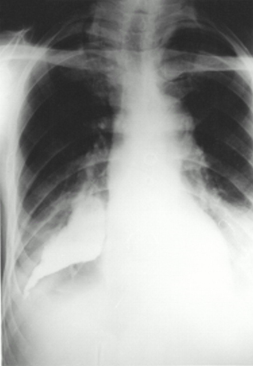
Five months after initiation of NIPPV the patient developed anorexia, weight loss of 4 kg, and lethargy. Serum inflammatory markers showed significant deterioration (CRP=150 mg/l). Assessment in hospital demonstrated a low grade fever after the application of NIPPV. Chest radiography revealed new opacification at the right lung base and the possibility of chronic pleural infection was raised. A further contrast swallow was arranged (fig 1) which showed the presence of an oesophagopleural fistula with leak of contrast into the right pleural space. NIPPV was discontinued. Surgery to repair the defect was considered but, given the profound respiratory muscle weakness and associated anaesthetic risk, this option was not pursued. The insertion of a PEG tube was offered but was declined by the patient. His health continued to decline with ongoing chronic sepsis. He died 4 months later.
Thin barium swallow demonstrating oesophagopleural fistula.
DISCUSSION
FSHMD is one of the more common inherited neuromuscular disorders with a prevalence of 1:20 000.1 We strongly suspected the diagnosis of FSHMD in our patient and, based on his phenotype, this was also the opinion of a consultant neurologist. The patient, however, declined formal diagnosis with electrophysiological studies and muscle biopsy. Symptomatic ventilatory failure is a rare but recognised feature of FSHMD2 and diaphragmatic weakness has been described.3 Nocturnal hypoventilation leading to daytime somnolence may be a prominent feature of primary muscle disorders, and nocturnal ventilatory support is a successful treatment.
Boerhaave’s syndrome is a rare but catastrophic condition with high mortality. The mechanism originally described by Herman Boerhaave in the 18th century is believed to be barogenic.4 The selection of patients for conservative or surgical approaches to management is not standardised.5 Recurrent leakage, even after surgical closure, has been described.6
NIPPV has an established role in the treatment of chronic ventilatory failure due to both neuromuscular and restrictive chest wall disease.7 It is known to increase mean oesophageal pressures,8 and gastric insufflation is a common but rarely troublesome complication occurring in up to 50% of patients.9 A review of the complications of NIPPV notes the possibility of pulmonary barotrauma and pneumothorax9 but there is, to our knowledge, no published account of oesophageal perforation.
The temporal relationship between the initiation of NIPPV and the onset of our patient’s anorectic illness suggests that this treatment had caused the radiologically healed oesophagopleural fistula to re-open. We believe this is the first report to describe re-opening of an oesophagopleural fistula secondary to the use of NIPPV. As the use of NIPPV becomes more widespread, this case provides a timely reminder to consider possible effects on the upper gastrointestinal tract, in addition to those on the respiratory system. We suggest that a previous history of oesophageal rupture be considered a relative contraindication to the initiation of NIPPV.
Acknowledgments
We are indebted to Dr A H Nickol for performing respiratory muscle studies on this patient.
Footnotes
-
Funding: none.
-
Conflicts of interest: none.