Thorax

HOME HELP FEEDBACK SUBSCRIPTIONS ARCHIVE SEARCH TABLE OF CONTENTS REGISTER
[Advanced]

Thorax 2007;62:652; doi:10.1136/thx.2006.074724
Copyright © 2007 BMJ Publishing Group Ltd & British Thoracic Society

This Article
Right arrow Extract Freely available
Right arrow Full Text (PDF)
Right arrow Alert me when this article is cited
Right arrow Alert me if a correction is posted
Services
Right arrow Email this link to a friend
Right arrow Similar articles in this journal
Right arrow Similar articles in PubMed
Right arrow Add article to my folders
Right arrow Download to citation manager
Right arrowRequest Permissions
Citing Articles
Right arrow Citing Articles via Google Scholar
Google Scholar
Right arrow Articles by Demura, Y
Right arrow Articles by Itoh, H
Right arrow Search for Related Content
PubMed
Right arrow PubMed Citation
Right arrow Articles by Demura, Y
Right arrow Articles by Itoh, H
Topic Collections
Right arrow Thorax Images in Thorax

IMAGES IN THORAX

Persistent diffuse pulmonary interstitial emphysema mimicking pulmonary emphysema

Y Demura, T Ishizaki, M Nakanishi, S Ameshima, H Itoh

Department of Respiratory Medicine, Faculty of Medical Sciences, University of Fukui Hospital, Fukui, Japan

Correspondence to:
Correspondence to:
Dr Y Demura
23-2 Matsuokashimoaizuki, Eiheiji-cho, Yoshida-gun, Fukui 910-1193, Japan; demura{at}fmsrsa.fukui-med.ac.jp

A 69-year-old male non-smoker with a history of atopic asthma of >30 years had recently been diagnosed with pulmonary emphysema (chronic obstructive pulmonary disease, COPD) because of exertional dyspnoea and chronic airflow limitation without bronchodilator responsiveness. A laboratory examination revealed a high serum IgE level (1219 U/ml) and a normal {alpha}1-antitrypsin level (142 mg/dl), and the flow-volume curve showed severe chronic airflow obstruction (vital capacity (VC) 2.47 litres (72.3% predicted), forced expiratory volume in 1 s (FEV1) 0.73 litres (27.4% predicted), FEV1%-Gaensler 30.8%). The chest radiograph showed severe hyperinflation (fig 1AGo), but carbon monoxide transfer factor was within normal limits (15.43 ml/min/mm Hg 98.9% predicted). A chest CT scan revealed the presence of free air predominantly along the bronchovascular sheaths of both lungs with thickened bronchial walls; pulmonary emphysema, pneumothorax and pneumomediastinum were not observed (fig 2AGo and B). The previous repeated episodes of asthma could be considered as the cause of the pulmonary interstitial emphysema. The patient was treated with inhaled cortico-steroids, long-acting ß2 agonists and leucotriene receptor antagonists. A few months later his respiratory symptoms had almost disappeared and the pulmonary function parameters had improved (VC 2.71 litres (78.9% predicted), FEV1 1.04 litres (40.3% predicted), FEV1.0%-G 40.6%). However, the CT findings remained unchanged throughout the 2 year observation period (fig 3Go).


Figure 1
View larger version (139K):
[in this window]
[in a new window]
[PowerPoint for Teaching]
 
Figure 1  Chest radiograph before treatment showing hyperinflated lungs and a decrease in pulmonary markings.


Figure 2
View larger version (28K):
[in this window]
[in a new window]
[PowerPoint for Teaching]
 
Figure 2  
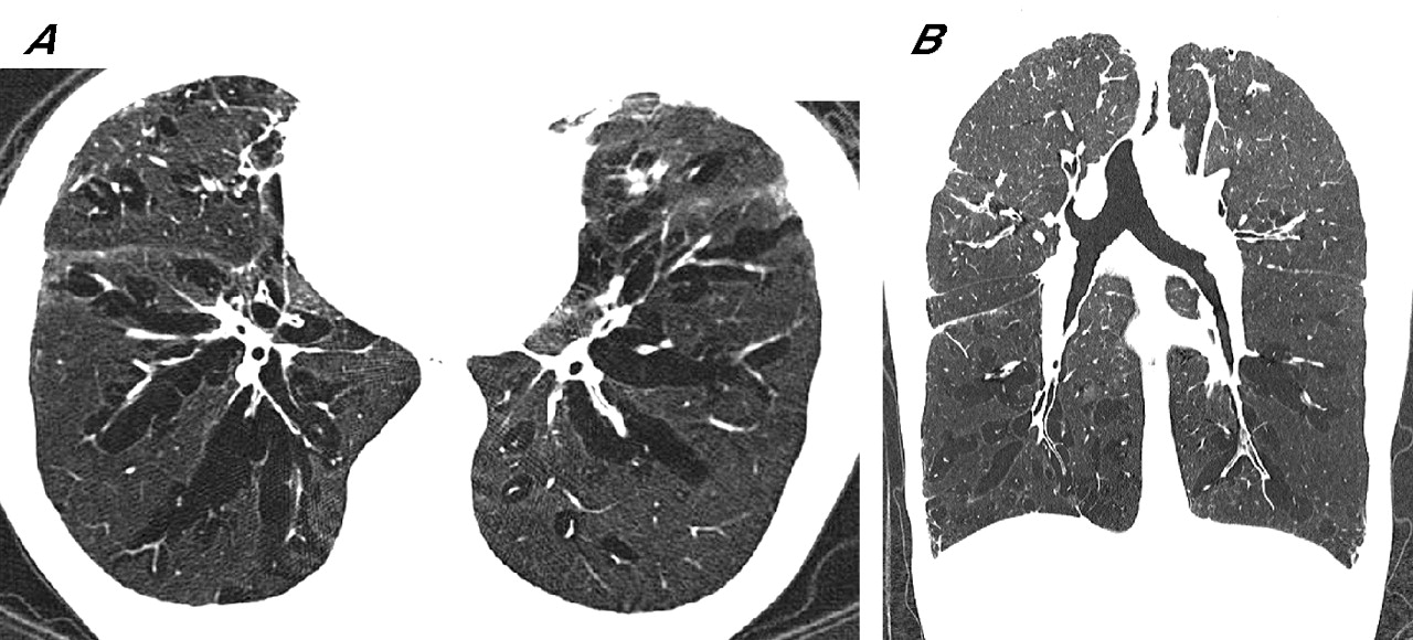
(A) Transaxial and (B) coronal sections of the chest CT scan before treatment showing diffuse pulmonary interstitial emphysema with multiple air lucencies contiguous to pulmonary vessels and bronchi on both lung fields.


Figure 3
View larger version (79K):
[in this window]
[in a new window]
[PowerPoint for Teaching]
 
Figure 3  Chest CT scan 2 years later still showing diffuse pulmonary interstitial emphysema after treatment.

 
Pulmonary interstitial emphysema has been reported infrequently, mostly in newborn infants,1 and is associated with the Macklin effect2 which is seen in blunt chest trauma, neonatal respiratory distress syndrome, asthma crises and positive pressure mechanical ventilation. To our knowledge, spontaneous diffuse pulmonary interstitial emphysema in association with persistent asthma has not previously been reported and could be confused with pulmonary emphysema.


Learning points

  • Diffuse pulmonary interstitial emphysema complicated by persistent asthma is extremely rare.
  • The high resolution CT findings are characteristic and should help in differentiating pulmonary interstitial emphysema from pulmonary emphysema (COPD).


References

  1. Jabra AA, Fishman EK, Shehata BM, et al. Localized persistent pulmonary interstitial emphysema: CT findings with radiographic-pathologic correlation. AJR Am J Roentgenol 1997;169:1381–4.[Free Full Text]
  2. Wintermark M, Schnyder P. The Macklin effect: a frequent etiology for pneumomediastinum in severe blunt chest trauma. Chest 2001;120:543–7.[CrossRef][Medline]


This Article
Right arrow Extract Freely available
Right arrow Full Text (PDF)
Right arrow Alert me when this article is cited
Right arrow Alert me if a correction is posted
Services
Right arrow Email this link to a friend
Right arrow Similar articles in this journal
Right arrow Similar articles in PubMed
Right arrow Add article to my folders
Right arrow Download to citation manager
Right arrowRequest Permissions
Citing Articles
Right arrow Citing Articles via Google Scholar
Google Scholar
Right arrow Articles by Demura, Y
Right arrow Articles by Itoh, H
Right arrow Search for Related Content
PubMed
Right arrow PubMed Citation
Right arrow Articles by Demura, Y
Right arrow Articles by Itoh, H
Topic Collections
Right arrow Thorax Images in Thorax


HOME HELP FEEDBACK SUBSCRIPTIONS ARCHIVE SEARCH TABLE OF CONTENTS REGISTER
Terms and conditions relating to subscriptions purchased online  ¦  Website terms and conditions  ¦  Privacy policy
Copyright © 2007 BMJ Publishing Group Ltd & British Thoracic Society