Article Text

Statistics from Altmetric.com
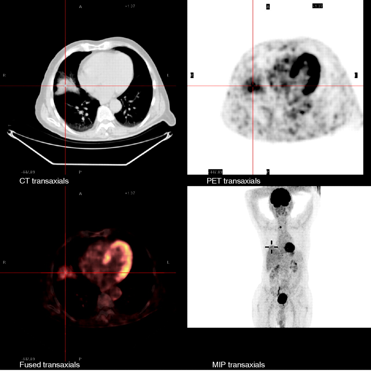
A 65-year-old man underwent lobectomy following a positron emission tomography (PET) scan showing increased uptake in a nodular lesion in the middle lobe (fig 1). A preoperative transbronchial biopsy specimen demonstrated lipoid pneumonia which was disregarded as a false positive (fig 2). No tumour was seen in the excised lobe.

Fluorodeoxyglucose positron emission tomography (PET) fusion CT scan showing moderately increased uptake in the middle lobe nodule (at the crossing point of the red lines). On CT scanning the lesion is approximately −50 Hounsfield units, consistent with fat.

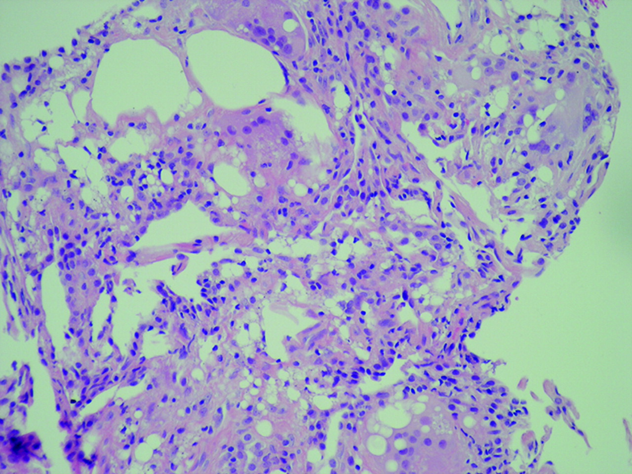
Transbronchial biopsy specimen showing alveolar inflammation, predominantly macrophages. Vacuoles are present within the alveoli and macrophages representing lipid material (black arrowheads). Giant cells also contain lipid (large black arrows). H&E stain; ×200 magnification.
Lipoid pneumonia is caused by aspiration of exogenous oils. It usually causes lung infiltrates but may also assume a nodular form suggesting a tumour.1 The PET scan may be “positive” as a result of metabolic activity of inflammation.2 Physicians should consider lipoid pneumonia in the differential diagnosis of a PET-positive lung lesion to avoid unnecessary surgery.
Learning points
-
Lipoid pneumonia may take a variety of forms, including mass lesions.
-
The history of oil ingestion is frequently not elicited until after the pathological diagnosis is made.
-
Inflammatory lung lesions may show increased uptake on PET scanning.
-
If a lung biopsy shows lipoid pneumonia in a PET-positive lesion, a conservative approach may be warranted.
Footnotes
-
Funding: None.
-
Competing interests: None.