
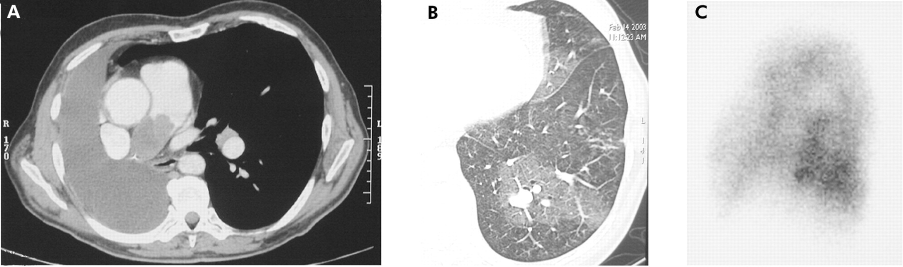
Figure 1
(A) Spiral CT scan after right pneumonectomy showing an endoluminal filling defect in the right pulmonary artery stump and the absence of additional thrombi in subsegmental branch arteries. (B) Transverse thin section CT scan showing multiple dilated vessels in the left lower lobe and mosaic areas of ground glass attenuation in the left lower lobe and the lingula. (C) Lung perfusion scan using 99mTc macroaggregated serum albumin showing multiple peripheral defects, particularly in the lingula and left Fowler segment (lateral view).
Vol 79 Issue 5
Table of Contents


Share this article
Click the icon of the social media platform on which you would like to share this article.


Email this article to a friend
Respond to this article