
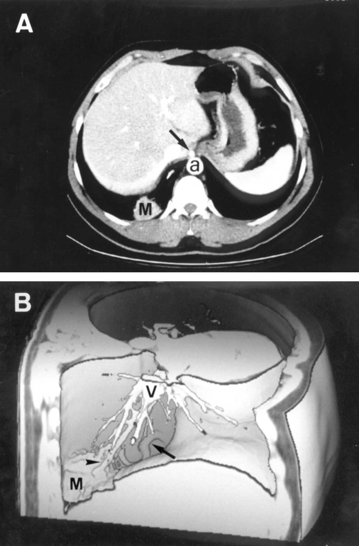
Figure 2
Spiral CT scan of case 2. (A) Contrast enhanced CT axial image showing the pulmonary mass (M) supplied by an anomalous systemic artery (arrow) arising from the upper abdominal aorta (a). (B) Right view of helical CT angiogram showing the course of the arterial vessel (arrow) to the pulmonary mass (M). Note the venous drainage (arrowhead) to the pulmonary veins (V).
Vol 79 Issue 5
Table of Contents


Share this article
Click the icon of the social media platform on which you would like to share this article.


Email this article to a friend
Respond to this article